Can brain cells recruit inflammation again after gastritis recovery? PVN ensembles and Hcn1
A mouse study follows hypothalamic cells recruited by gastritis and finds that post-recovery reactivation still alters gastric state, linking neural, endocrine and channel-related clues.
Gastric inflammation signals to the brain, but how can the brain’s response influence the stomach? A 2026 Science Advances study labels PVN cells recruited during chemical gastritis in mice and examines their function after gastric tissue recovers. Reactivating these cells reintroduces local gastric abnormalities. The work follows neural and endocrine outputs and identifies a channel-related Hcn1 lead, connecting post-recovery brain activity with organ state.
Gastritis-recruited PVN ensembles and their DMV output regulate gastric inflammation. Repeated reactivation after recovery can reinduce local abnormalities, stress recruits related cells, and local Hcn1 inhibition attenuates outcomes.
Contents · 6 sections
Inflammation recruits brain cell ensembles
Bidirectional brain–stomach communication makes inflammation both a peripheral event and a state that recruits central responses. Identifying participating cells allows a distinction between activity accompanying inflammation and activity capable of changing it. Following the same ensemble after recovery is an important extension of this approach.
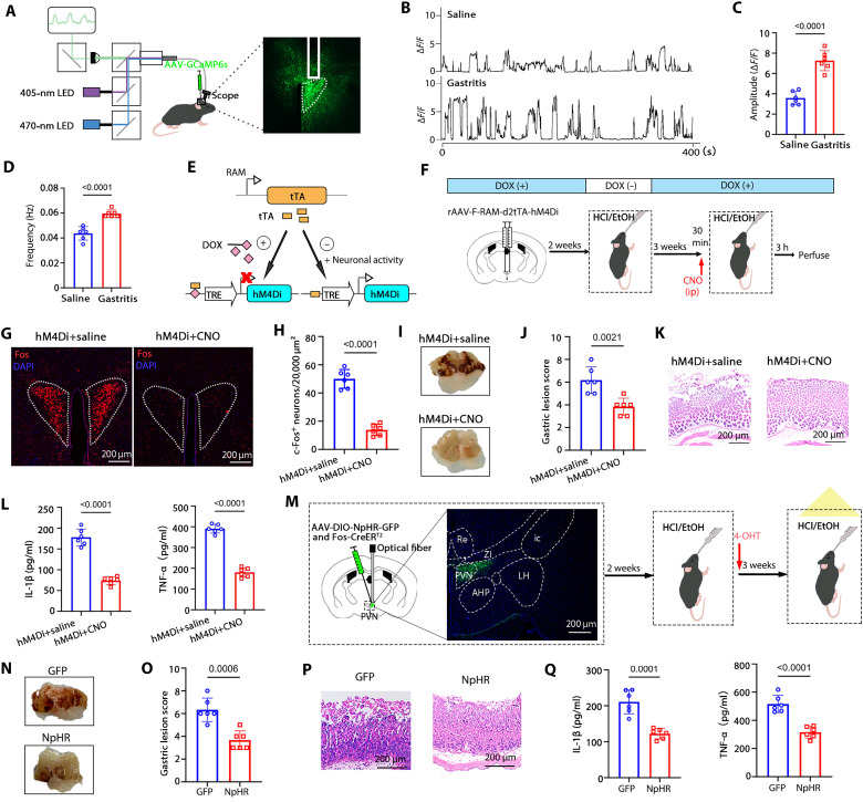
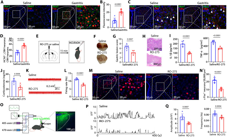
Chemical gastritis produced gastric hemorrhage, architectural abnormalities and elevated local cytokines. Gastric tracing combined with activity labeling identified related cells in several regions, with prominent overlap in PVN and increased regional calcium activity. Gastric state was thus connected with observable brain responses.
Suppressing cells labeled during the initial inflammation attenuated a later challenge after recovery. Optical and chemogenetic strategies converged, and PSTh suppression also had a smaller effect. Activity-defined ensembles recruited during inflammation could therefore participate in later organ regulation within a broader network.
Source: Liu E et al. · Science Advances · CC BY-NC
Original figure and English caption ↗
Neural and endocrine outputs show distinct responses
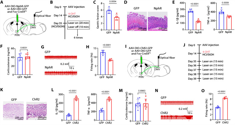
The labeled ensemble partly overlapped CRH cells; gastritis accompanied higher corticosterone, and CRH-receptor blockade attenuated inflammation. Tracing, terminal stimulation and gastric nerve firing supported a PVN→DMV→stomach connection. Vagotomy only partly reduced PVN activity, while cytokines could also recruit cells, indicating multiple incoming routes.
Suppressing the relevant DMV terminals reduced inflammation and gastric firing while corticosterone remained similar. Terminal activation induced mild local inflammation without significant corticosterone or serum-cytokine elevation. Different local and systemic responses clarified the distinction between neural and endocrine components.
Source: Liu E et al. · Science Advances · CC BY-NC
Original figure and English caption ↗
Previously recruited cells retain influence over the stomach
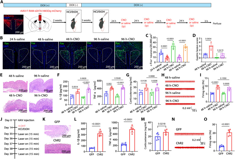
Repeated ensemble enhancement sustained gastric abnormalities and firing-related changes. After tissue recovery, several days of optical reactivation reintroduced local structural and cytokine abnormalities without parallel serum-cytokine increases. Previously recruited brain cells retained the capacity to influence the organ after recovery.
The result extended the investigation from an immediate brain–stomach response to a regulatory state accessible after recovery. Local responses to repeated stimulation made the cells an entry point for studying persistent changes. Their recruitment during natural challenges became a question connecting these observations with stress conditions.
Source: Liu E et al. · Science Advances · CC BY-NC
Original figure and English caption ↗
Hcn1 relates to activity and inflammatory modulation
Single-nucleus profiling identified Hcn1 and CRH changes in inflammation-associated Fos-positive cells, with partial co-expression. Local Hcn1 inhibition reduced PVN activity, corticosterone and gastric nerve firing while attenuating gastritis. Molecular findings and regional function aligned, providing a channel-related lead for the influence of brain activity on the organ.
Partial Hcn1–CRH overlap also indicated diversity within the activity-defined population. Resolving channel roles in different cells and changes during inflammation, recovery and rechallenge will help explain how local excitability participates in longer-term regulation.
Source: Liu E et al. · Science Advances · CC BY-NC
Original figure and English caption ↗
Different challenges recruit related ensembles
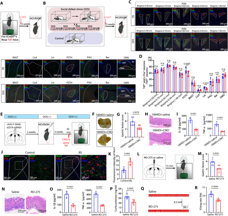
Cells recruited by water-immersion restraint and social defeat overlapped by about eighty percent with the prior gastritis-labeled ensemble. Suppression during restraint attenuated gastric abnormalities. Stress also accompanied higher Hcn1, and local inhibition improved related outcomes. Different challenges thus accessed partly shared cellular activity.
From initial gastritis to stress challenges, the relationship between brain ensembles and peripheral responses could be followed repeatedly. Shared activity, neural outputs and molecular modulation converged to make the influence of stress on gastric state a more defined circuit question.
Source: Liu E et al. · Science Advances · CC BY-NC
Original figure and English caption ↗
Following brain state after organ recovery
The work brings peripheral inflammation, central recruitment, and neural and endocrine outputs into one framework. Gastric tissue recovery does not eliminate the regulatory capacity of associated brain cells. Following these populations reveals relationships between organ state and brain activity across timescales.
The findings come from chemical gastritis and defined stress models in male mice. Other inflammatory causes, cell types and natural recovery processes remain directions for investigation. The results add defined cellular and molecular leads to post-recovery brain–stomach relationships, extending studies of persistent states beyond visible tissue recovery.