Mechanosensory nerves contribute to bone–implant integration
A mouse study links reduced mechanical loading with sensory signaling: Piezo2 neurons, CGRP and FAK connect to bone formation, alongside mechanical sensitivity and avoidance.
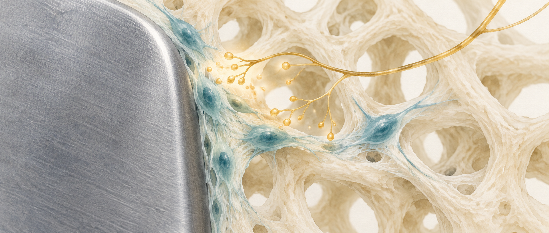
An orthopedic implant needs both load-bearing material and integration with surrounding bone. If metal bears more of the load, bone cells may receive less mechanical input—a phenomenon termed stress shielding. Could signals from bone-innervating nerves provide another approach alongside material changes? A study published in Nature Communications in 2025 follows a neuro-bone connection in male mice with tibial implants: neuropeptides from mechanosensory cells influence signaling and differentiation in osteogenic precursors.
Activating Piezo2-marked sensory cells increases peri-implant CGRP and early osseointegration measures. Culture and blockade experiments support involvement of CGRP–PKA–FAK signaling, while stimulation also produces mechanical hypersensitivity and place avoidance.
Contents · 7 sections
The same alloy with different stiffness elicits different bone responses
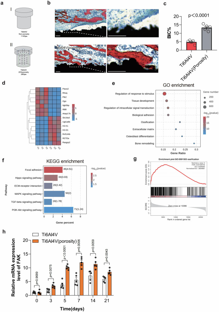
The team compares implants made from the same titanium alloy: a higher-modulus solid structure and a lower-modulus internally porous structure. After two weeks, bone contact is lower around solid implants, alongside differences in osteogenesis-related transcription. Material load bearing is thus associated with local tissue responses.
Differences in focal adhesion kinase, FAK, highlight mechanical signaling. FAK links cells to their surrounding matrix and helps translate external influences into intracellular responses. Solid implants show lower FAK expression and a later peak, suggesting less timely recruitment of signaling involved in bone formation.
Source: Wang Q et al. · Nature communications · CC BY-NC-ND
Original figure and English caption ↗
Bone mechanical information also involves sensory nerves
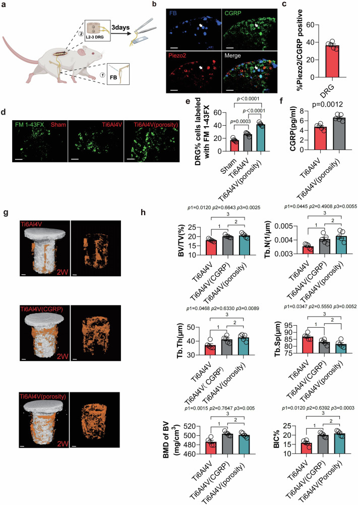
Tracing from the tibia identifies lumbar dorsal-root-ganglion sensory cells, some bearing both mechanosensory-related Piezo2 and calcitonin gene-related peptide, CGRP. Higher-modulus implants accompany lower neuronal-activation readouts and lower peri-implant bone CGRP, connecting material differences with neuropeptide signaling.
Reducing local Piezo2-marked cells lowers CGRP around both implant types, whereas reducing other tested populations does not produce the same result. Local CGRP supplementation increases bone volume, bone contact and FAK expression around solid implants, showing that the peptide can influence this environment.
Source: Wang Q et al. · Nature communications · CC BY-NC-ND
Original figure and English caption ↗
Increasing neuropeptides also changes sensory behavior
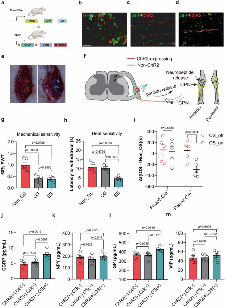
The team expresses a light-sensitive protein in Piezo2-marked cells and stimulates dorsal-root ganglia with a small wireless implanted light source. CGRP and substance P increase, while two other measured neuropeptides do not change significantly, indicating selective changes in signaling.
Stimulation also has sensory consequences: mechanical withdrawal thresholds fall and mice avoid the stimulation-paired location. Thermal sensitivity does not increase significantly, whereas the electrical-stimulation comparison does increase thermal sensitivity. Cell selection changes the response profile, but bone-integration benefits still accompany sensory changes.
Source: Wang Q et al. · Nature communications · CC BY-NC-ND
Original figure and English caption ↗
Recruiting sensory cells increases bone–material contact
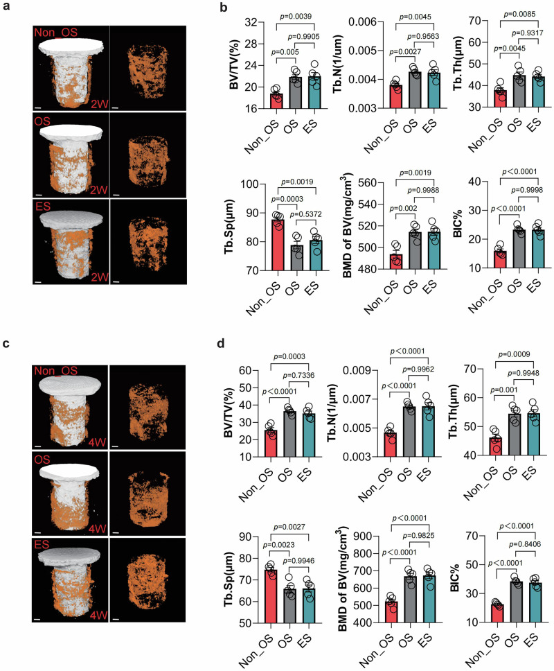
At two and four weeks after implantation, micro-CT shows increased peri-implant bone-volume fraction, trabecular number and thickness, mineral density and bone-contact proportion, with reduced trabecular separation. Electrical stimulation produces changes in the same direction; optical stimulation is not uniformly superior in these comparisons.
The changes extend the neuropeptide findings to tissue. Around the same class of higher-modulus implant, more bone and greater contact develop. The approach concerns altering local biological responses alongside the mechanical load-bearing properties of the material.
Source: Wang Q et al. · Nature communications · CC BY-NC-ND
Original figure and English caption ↗
New bone accompanies changes in CGRP
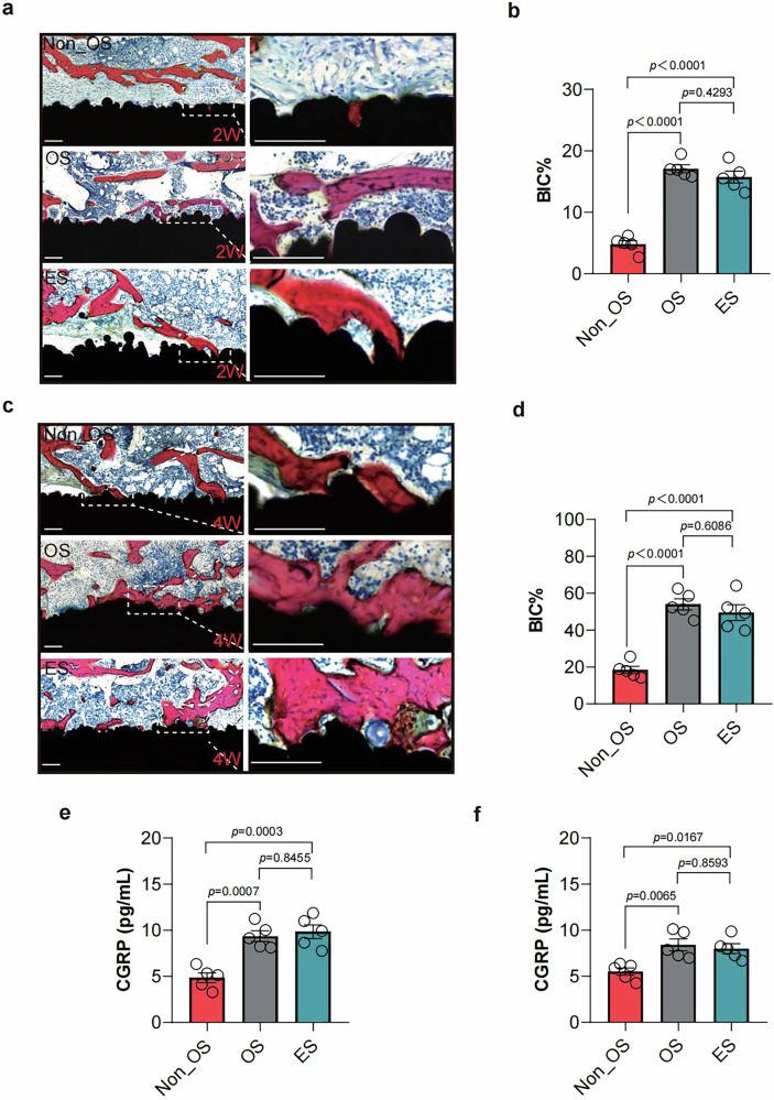
Hard-tissue sections likewise show more new bone near stimulated implants and greater contact. Peri-implant CGRP is higher at both observation times, while osteogenic markers and collagen-related observations further support increased local bone formation.
Inflammatory readouts do not show a broad decline: IL-10 differs, while IL-1β, IL-6 and TGF-β do not change significantly. Relative to these limited inflammatory changes, the link between neuropeptides, osteogenesis and bone contact leads more directly to investigation of intracellular signaling in bone-related cells.
Source: Wang Q et al. · Nature communications · CC BY-NC-ND
Original figure and English caption ↗
Neural conditioned medium changes osteogenic-precursor responses
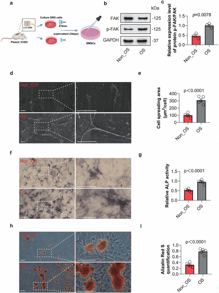
Conditioned medium from optically stimulated ganglion cells increases FAK phosphorylation in bone-marrow mesenchymal stromal cells. With titanium alloy, cell-spreading area increases; alkaline-phosphatase activity and mineralized nodules also increase in osteogenic culture. Soluble neural signals thus influence another cell class.
An osteoclast-differentiation experiment does not show a corresponding significant change. The findings therefore emphasize osteogenesis, combining altered cell morphology at the material surface with differentiation and mineralization measurements.
Source: Wang Q et al. · Nature communications · CC BY-NC-ND
Original figure and English caption ↗
CGRP–PKA–FAK links neural signaling with bone
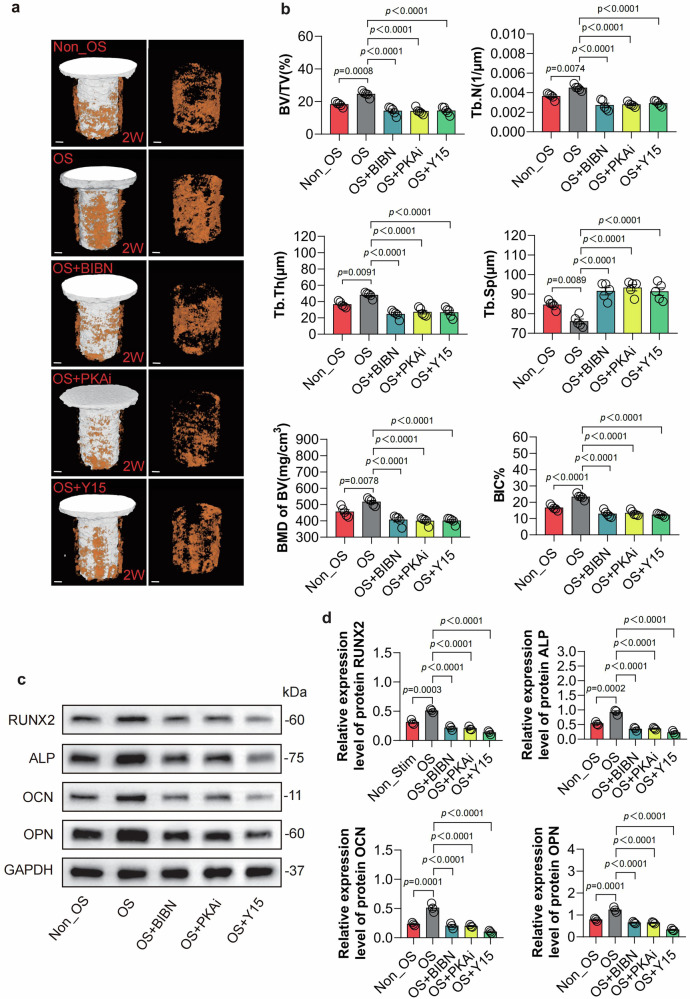
Blocking CGRP receptors or inhibiting PKA or FAK attenuates stimulation-associated increases in osseointegration and osteogenic markers. Substance-P receptor blockade does not clearly suppress the improvement, and light alone in ordinary mice does not produce the same benefit. These contrasts strengthen the involvement of CGRP-related signaling.
This adult male mouse study shows that peri-implant bone responses are influenced by sensory-neural signals. Endogenous neuropeptides can accompany greater osteogenesis in a mechanically less stimulated environment. The evidence centers on early tissue integration; these observations do not directly establish long-term load-bearing performance or loosening risk.
Source: Wang Q et al. · Nature communications · CC BY-NC-ND
Original figure and English caption ↗