How the brainstem divides the work of making less heat and losing more
Two parabrachial cell populations defend mice against warming through different emphases: suppressing brown-fat heat production and increasing heat loss through the tail.
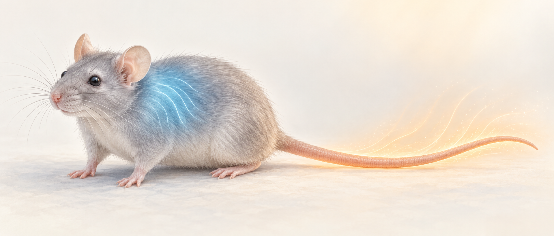
When the environment warms, the body can turn down its internal furnace and open routes for heat loss. Are these jobs assigned together, or separated earlier in the brain? A mouse study published in Science Advances on September 2, 2020 examines the lateral parabrachial nucleus (LPB) in the brainstem. Two populations projecting toward the hypothalamic preoptic area both help lower body temperature, yet influence different components of cooling.
Pdyn-marked LPB inputs preferentially suppress brown-fat heat production, whereas CCK-marked inputs promote tail heat loss. Both also influence shivering, revealing distinct but overlapping components of heat defense.
Contents · 7 sections
Cooling has two levers: make less heat and lose more
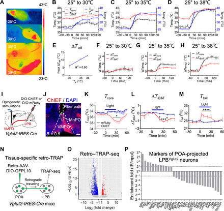
Body temperature is not a simple reflection of the surroundings. Brown fat consumes energy to produce heat, while skin blood flow carries heat out of the body. The tail is an important heat-loss surface in mice. As ambient temperature rises, tail temperature increases and brown-fat heat production is suppressed, shifting the balance from keeping warm to defending against heat.
LPB conveys body-temperature information toward the preoptic area. Increasing its glutamatergic input to the ventromedial preoptic area (VMPO) lowers core and brown-fat temperatures while raising tail temperature. These coordinated changes raise a question: does this common route already contain lanes assigned to different cooling jobs?
Source: Yang WZ et al. · Science advances · CC BY-NC
Original figure and English caption ↗
Two populations respond to warmth
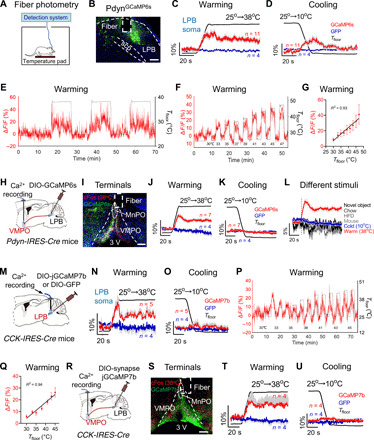
The team identifies LPB populations marked by Pdyn and CCK. Most are glutamatergic, and the two populations overlap little. Warmth recruits both, but differently: the CCK population accounts for a larger fraction of heat-responsive cells as warming becomes stronger.
Calcium recordings show warmth responses in both populations and their VMPO terminals, with larger responses to stronger warming. Pdyn somata also show a small transient response to cooling, whereas the cooling response at their VMPO terminals is not significant. Temperature signals therefore differ across populations and projection sites.
Source: Yang WZ et al. · Science advances · CC BY-NC
Original figure and English caption ↗
A shared destination, different jobs
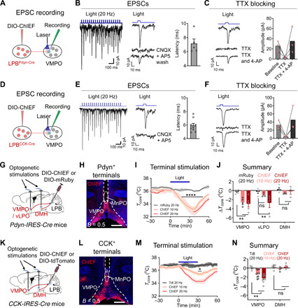
Both populations make direct excitatory connections to VMPO, and increasing either VMPO input lowers core temperature. Corresponding inputs to the dorsomedial hypothalamus do not significantly change core temperature under the tested conditions. The destination of the signal matters.
Judged only by the drop in core temperature, the two inputs seem to perform the same task. Following brown fat, the tail and shivering reveals their different emphases. The thermometer reports the overall balance; changes in individual organs show how heat production is reduced or heat loss increased.
Source: Yang WZ et al. · Science advances · CC BY-NC
Original figure and English caption ↗
The Pdyn branch turns down the brown-fat furnace
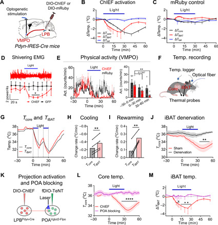
Increasing Pdyn-marked LPB input to VMPO lowers brown-fat temperature before core temperature, with a faster and larger decline. Tail temperature does not rise significantly. This timing fits reduced internal heat production. Shivering is also partly reduced, diminishing another source of heat.
When brown fat lacks sympathetic innervation, this input no longer produces cooling. Reducing activity in the preoptic glutamatergic population likewise prevents the falls in core and brown-fat temperatures. Together, these findings connect this branch with preoptic control of brown-fat heat production.
Source: Yang WZ et al. · Science advances · CC BY-NC
Original figure and English caption ↗
The CCK branch opens a route for tail heat loss
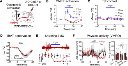
CCK-marked input produces a different picture: the tail warms, core temperature falls and shivering is suppressed. Brown-fat temperature also declines, but approximately alongside core temperature, consistent with following whole-body cooling rather than leading it through reduced heat production.
CCK input can still produce cooling when brown fat lacks sympathetic innervation. This contrasts with Pdyn input: one branch primarily reduces heat production, while the other favors heat loss. Their shared influence on shivering also shows that these jobs are not completely separate.
Source: Yang WZ et al. · Science advances · CC BY-NC
Original figure and English caption ↗
Without the heat-production brake, warmth is harder to manage
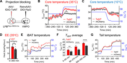
The researchers next reduce activity in Pdyn cells projecting to the preoptic area. In warmth, core temperature, energy expenditure and brown-fat temperature increase; core temperature in the cold does not change significantly. Locomotion remains broadly stable across the tested environments, so the thermal changes do not accompany a general increase in movement.
Tail temperature rises later during warmth, potentially compensating for the higher core temperature. Heat loss can still increase, yet does not fully offset the burden of excess heat production. The Pdyn population thus contributes to ordinary heat defense beyond simply being capable of producing cooling.
Source: Yang WZ et al. · Science advances · CC BY-NC
Original figure and English caption ↗
Two cooling routes connect temperature with energy balance
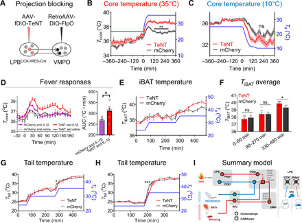
Reducing activity in preoptic-projecting CCK cells delays tail warming and promotes heat accumulation in warm surroundings, while cold core temperature remains without a significant change. Reducing this population also worsens inflammatory fever, linking control of heat loss to fever severity.
A separate experiment involving the broader Pdyn population connects heat production with energy balance: reducing its activity increases energy expenditure and reduces weight gain during high-fat feeding, without significant changes in food intake or locomotion. The molecular markers identify this division of labor, while the roles of the peptides and downstream cell types remain unresolved. These mouse findings reveal heat defense as coordinated work across several components and give a concrete connection to metabolism.
Source: Yang WZ et al. · Science advances · CC BY-NC
Original figure and English caption ↗