How can a gut metabolite influence compulsive-like behavior? Two pathways behind succinate-associated changes
The study follows succinate through prefrontal microglia, vagal signaling and behavior, examining different routes by which metabolic signals affect brain circuits.
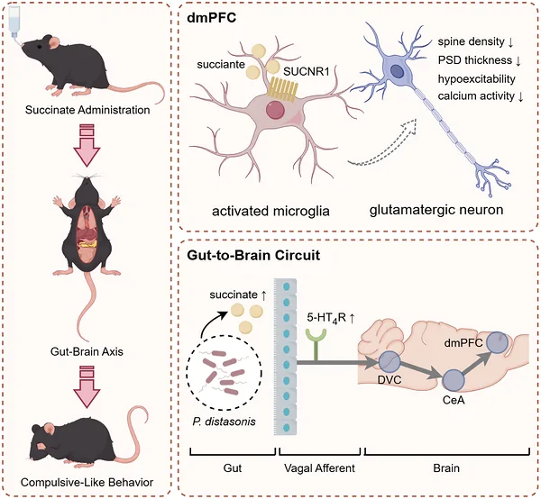
Why might a small molecule in the gut make mice groom repeatedly? Succinate is a familiar component of metabolism, yet this study traced its effects through brain immune cells, the vagus nerve and prefrontal neurons. Following two routes, the researchers turned the broad idea of gut–brain communication into a more specific circuit story.
Succinate-associated compulsive-like behavior was linked to reduced activity of prefrontal glutamatergic neurons. Microglial SUCNR1 and vagus-associated signaling provided two experimentally testable mechanistic pathways.
Contents · 7 sections
The story begins with an elevated metabolite
Intrusive thoughts and repetitive behavior are characteristic features of obsessive-compulsive disorder. In mice, excessive grooming offers a tractable behavioral window into the biology of repetition. The study asks how gut-associated metabolic changes might contribute to this compulsive-like pattern.
Earlier work by the team linked patient-derived microbiota transplantation in mice with behavioral changes and elevated succinate. This Advanced Science study, published in 2026, asks whether succinate is merely an associated marker or participates through identifiable cells and pathways.
What does repeated grooming reveal?
After succinate exposure, mice showed more grooming bouts and longer grooming following water spray. Weight, water intake, overall movement and selected anxiety-related measures did not differ significantly. Spontaneous behavior also shifted toward grooming and away from exploration, indicating a change in behavioral allocation rather than simply increased movement.
The researchers also examined the succinate-producing bacterium P. distasonis. Colonized mice showed both anxiety-like and compulsive-like changes, a pattern that differed from succinate exposure alone. This distinction points to additional microbial signals while making succinate a useful entry point for further investigation.
Why did prefrontal activity fall?
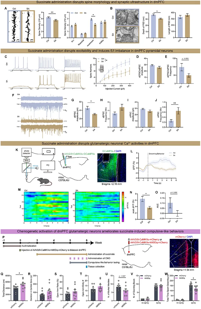
Succinate exposure was accompanied by fewer mature dendritic spines, thinner postsynaptic densities and altered excitatory–inhibitory input in the dmPFC. Glutamatergic neurons also showed weaker grooming-related calcium responses. Structural, electrophysiological and in vivo observations converged on the same local circuit node.
The team then increased the activity of these prefrontal glutamatergic neurons. Excessive grooming associated with succinate was reduced. The intervention connected structural and activity observations with behavior: changing the neuronal state changed the repetitive-grooming phenotype, giving the gut-derived signal a defined brain target.
Source: Li Y et al. · Advanced science (Weinheim, Baden-Wurttemberg, Germany) · CC BY
Original figure and English caption ↗
Microglia pick up the succinate signal
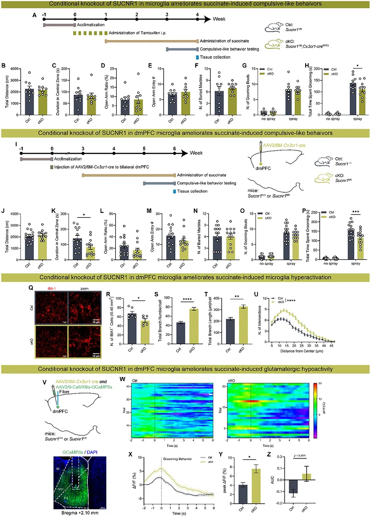
A second line of evidence focused on microglia. Succinate exposure changed their abundance, morphology and transcriptional state in the dmPFC. The authors proposed that the succinate receptor SUCNR1 could translate a metabolic change into an immune-cell response within the brain.
Conditional removal of SUCNR1 from microglia reduced excessive grooming, improved selected morphological changes and restored behavior-related calcium signals in prefrontal glutamatergic neurons. Behavior, immune cells and neurons responded in the same direction. SUCNR1 emerged as a link between metabolic changes and the brain state, leaving the precise microglia-to-neuron signal as the next question.
Source: Li Y et al. · Advanced science (Weinheim, Baden-Wurttemberg, Germany) · CC BY
Original figure and English caption ↗
The vagus nerve offers another route to the brain
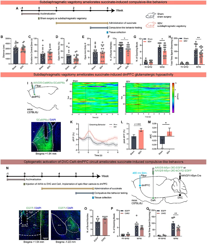
Gut-derived information may also reach the brain through neural signaling. Vagal interruption reduced compulsive-like behavior and increased dmPFC glutamatergic calcium activity. Tracing and pathway interventions supported a model involving the dorsal vagal complex, central amygdala and prefrontal cortex.
Gut-associated 5-HT4 receptors provided another clue. Their expression changes were linked to behavioral measures, while receptor antagonism reduced the behavioral abnormalities and improved prefrontal neuronal signals. These findings connect succinate, intestinal receptor signaling and vagal input, opening questions about how metabolic signals alter receptors and reach the brain.
Source: Li Y et al. · Advanced science (Weinheim, Baden-Wurttemberg, Germany) · CC BY
Original figure and English caption ↗
From animal models to clues in people
The team extended the investigation to Hoxb8-deficient mice, a model with compulsive-like behavior. Local inhibition of succinate dehydrogenase improved selected anxiety-like and compulsive-like measures alongside changes in neuronal activity labeling and microglial indices. The findings in this additional model strengthen interest in the metabolic link to brain-cell states.
In the human analysis, serum succinate was associated with selected prefrontal imaging measures and yielded an ROC area of 0.73 for distinguishing patients from healthy controls. Animal experiments describe cells and circuits, while these observations add a human association. Larger samples and independent validation will be needed to assess succinate as a candidate biomarker.
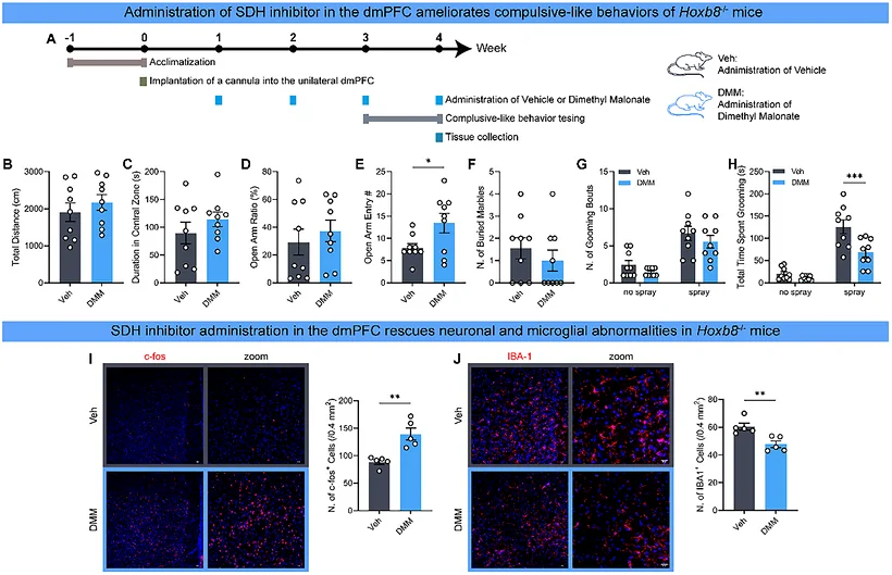
Source: Li Y et al. · Advanced science (Weinheim, Baden-Wurttemberg, Germany) · CC BY
Original figure and English caption ↗
Two routes converge on a prefrontal node
The work divides the metabolite–behavior relationship into two routes: one involving microglia and a receptor within the brain, and another involving peripheral neural signaling and central relays. Both converge on prefrontal glutamatergic state, providing a more specific framework than a microbial association alone.
The findings raise new questions: do the routes reinforce one another, which acts first, and how do local inhibitory cells join the conversation? Mechanistic evidence in male mice and associations in people offer complementary starting points. Following one small metabolite makes the connections among the gut, immune cells and neural circuits more tangible.