How the brain regulates movement when the stomach expands
Gastric dilation recruits hypothalamic CRH-related cells in male mice. Manipulating their activity and brainstem terminals changes gastric-movement amplitude; human brain-connectivity and gastric-electrical observations offer another clue.

The stomach is more than a food container: its relaxation, contraction and feedback involve continuous communication with the brain. How does expansion shape its subsequent movement? A study published in Frontiers in Neuroscience on February 22, 2023 combines brain imaging after water loading in healthy participants with cellular and circuit experiments in a male-mouse gastric-dilation model. It focuses on corticotropin-releasing-hormone-related (CRH) neurons in hypothalamic paraventricular nucleus (PVN) and their output toward the dorsal motor nucleus of the vagus (DMV). Dilation accompanies increased activity in selected cells, and reducing related activity increases gastric-movement amplitude.
Bidirectional population and local DMV-terminal manipulations support a role for PVN CRH-related output in gastric-movement regulation. Electroacupuncture-associated changes suggest a brain–body mechanism for further testing.
Contents · 6 sections
Brain and stomach change together in healthy participants
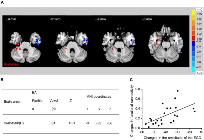
The study first examines 24 healthy young adults, with 12 men and 12 women. After water loading produces fullness, participants receive electroacupuncture, with resting-state imaging and gastric-electrical recordings compared before and after. Hypothalamic–brainstem functional connectivity increases, and its change correlates positively with gastric-electrical amplitude changes.
This within-group before-and-after observation links regional brain signals with gastric-electrical changes. Functional connectivity describes temporal signal correlation, and gastric electrical activity differs from stomach-wall contraction force. These observations direct attention to hypothalamus and brainstem, which mouse experiments then examine at cellular resolution.
Source: Wang XY et al. · Frontiers in neuroscience · CC BY
Original figure and English caption ↗
Gastric dilation recruits a PVN population
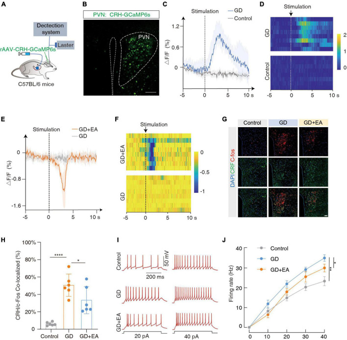
In male mice, gastric dilation rapidly increases calcium signals in a CRH-related PVN population. Tissue analysis shows greater CRH/c-Fos colabeling, while slice recordings reveal more action potentials under the same current drive. Calcium, activity markers and excitability provide complementary observations.
After electroacupuncture, population calcium, colabeling and current-evoked firing decrease in model mice. Both gastric state and somatic stimulation thus accompany altered activity in this population. The next question is whether these cells merely track bodily changes or can also influence gastric movement.
Source: Wang XY et al. · Frontiers in neuroscience · CC BY
Original figure and English caption ↗
Lowering or raising activity shifts gastric movement oppositely
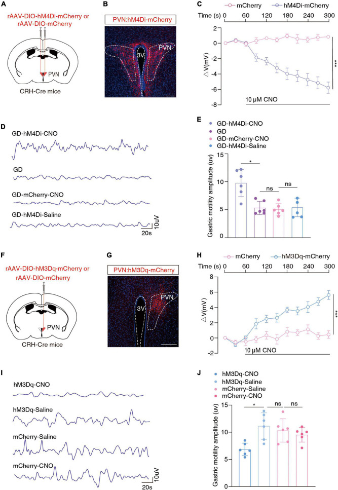
In dilation-model mice, inhibiting PVN CRH cells increases gastric-movement amplitude, unlike marker controls or conditions without activation of inhibition. Conversely, activating these cells in normal mice decreases amplitude. Bidirectional experiments show that population activity can alter a bodily measure beyond accompanying it.
Mouse movement is recorded with a stomach-wall sensor rather than the participants’ gastric-electrical measurement. A larger amplitude describes stronger measured gastric movement, while emptying and digestive efficiency are bodily functions requiring separate measurements. This physiological response motivates following the output route to a relay.
Source: Wang XY et al. · Frontiers in neuroscience · CC BY
Original figure and English caption ↗
Finding inhibitory input at a vagal output center
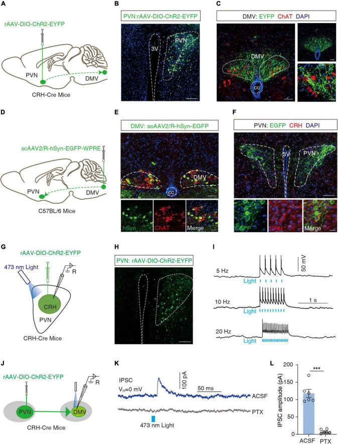
DMV is a brainstem center regulating gastric and other visceral functions, with many cholinergic cells. Anterograde tracing finds PVN CRH-cell fibers near cholinergic DMV cells, and retrograde tracing from DMV labels CRH-colabeled PVN cells. These observations map a connection between the regions.
Activating labeled PVN fibers in DMV slices evokes inhibitory postsynaptic currents in DMV cells; GABA-receptor blockade markedly reduces them. This supports an inhibitory functional connection. CRH-associated cell identity does not by itself identify CRH peptide as the cause of that current.
Source: Wang XY et al. · Frontiers in neuroscience · CC BY
Original figure and English caption ↗
Local terminals link brainstem input to gastric movement
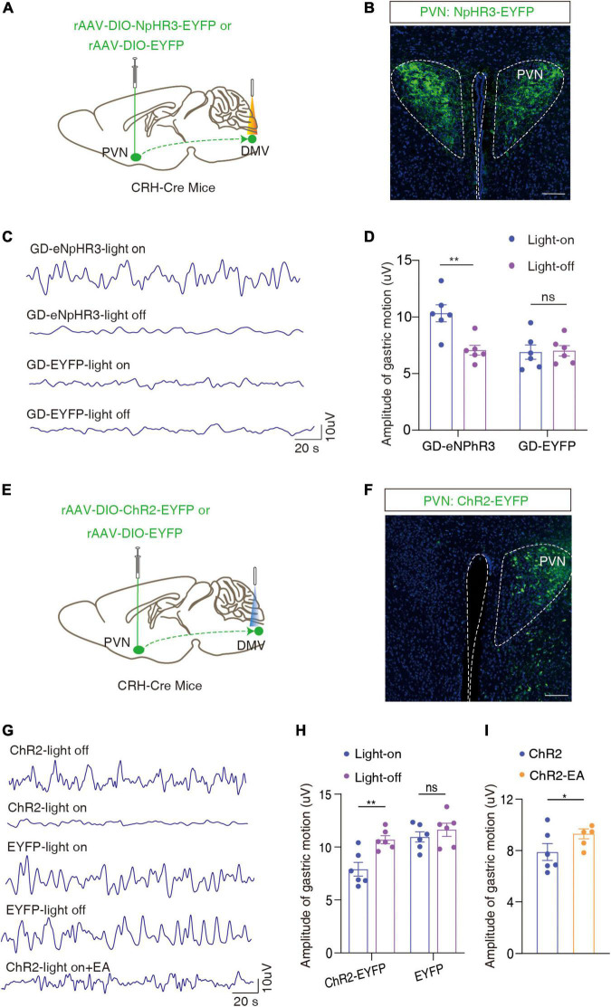
Restricting manipulation to PVN CRH-related terminals in DMV increases gastric-movement amplitude with inhibition in model mice and decreases it with activation in normal mice. Concordant directions at the population and local-output levels support a contribution to gastric regulation.
After terminal activation lowers amplitude, electroacupuncture still accompanies an increase. The authors connect this with reduced PVN activity and propose modulation of this output as a contributor. The experiments do not isolate the route as the sole or required mediator of electroacupuncture; other brain–body inputs may contribute.
Source: Wang XY et al. · Frontiers in neuroscience · CC BY
Original figure and English caption ↗
Gastric feedback meets central regulation
From cellular responses during dilation to hypothalamic output and local brainstem manipulation, the study connects bodily state and central regulation in an observable physiological process. Its central finding is that gastric-movement amplitude changes with selected central cells and inputs, illustrating regulation returning from brain to body.
Circuit experiments chiefly use 8–10-week-old male mice in a gastric-dilation model; the human component is a short-term before-and-after observation in healthy young adults. They provide cellular functional evidence and regional correlations, respectively, without testing efficacy in chronic digestive-disease patients. Together they clarify both mechanisms and the bodily states studied.