How does stress slow esophageal transit? Clues from a brain–vagal connection
A mouse study follows hypothalamic and vagal outputs into gastroesophageal molecules, finding distinct regulation of esophageal function and stress-related behavior.
Reduced feeding under stress can involve motivation or difficulty moving food through the esophagus. How can these contributions be distinguished? A 2026 study in mice exposed to chronic unpredictable mild stress, or CUMS, follows esophageal transit and sphincter pressure into hypothalamic PVN and dorsal motor vagal nucleus, or DMV, circuits, then into molecules at the gastroesophageal junction, or GEJ. A notable finding is that modulation of a defined brain–organ output improves esophageal function and feeding without simultaneously restoring stress-related behavior.
PVN–DMV–vagal and cholinergic signaling contribute to stress-associated esophageal changes. Reducing local MYBPC2 improves sphincter pressure and transit time during sustained pathway activation.
Contents · 6 sections
The passage of food through the esophagus changes
After four weeks of CUMS, daily feeding, weight gain and food-zone time decreased, with changes in exploration and coping tasks. Overall open-field distance and fasting-refeeding measures remained similar, while heart rate, blood pressure and stress hormones rose. Different feeding outcomes showed the influence of animal state and feeding conditions.
Esophageal measures provided more direct organ-level clues: transit time lengthened and lower esophageal sphincter pressure, or LESP, rose, while width remained similar. Epithelial and keratin layers thickened and GEJ inflammatory markers increased. Changes in passage and tissue state prompted investigation of outputs connecting the brain with the esophagus.

Source: Zhang K et al. · Molecular metabolism · CC BY-NC-ND
Original figure and English caption ↗
Tracing from the esophagus to PVN and DMV
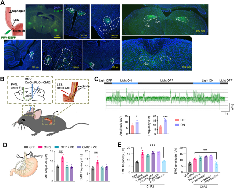
Organ-linked retrograde trans-synaptic tracing labeled several brain regions, with subsequent focus on PVN and DMV. Increasing the activity of DMV cells associated with PVN input and the peripheral target enhanced esophageal EMG. The rapid response disappeared after denervation, linking it with neural output.
Cholinergic antagonism also blocked the EMG enhancement, pointing to acetylcholine, or ACh, signaling. Anatomical, EMG and pharmacological findings converged on a modifiable brain–organ connection. Following it allowed comparison of whether organ function and stress-related behavior always change together.
Source: Zhang K et al. · Molecular metabolism · CC BY-NC-ND
Original figure and English caption ↗
Esophageal improvement with persistent stress-related behavior
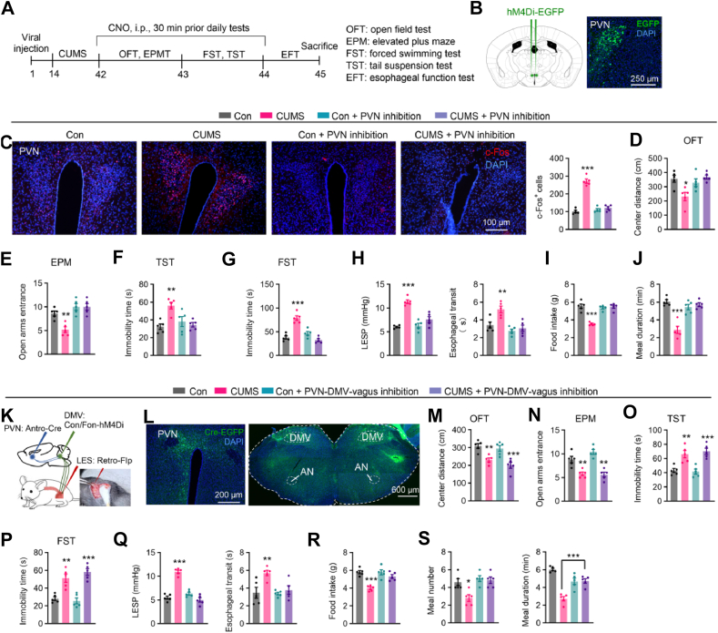
Esophageal vagal denervation improved transit time, LESP and tissue changes in stressed mice, with no additional benefit from cholinergic antagonism. Broad PVN inhibition improved esophageal function, feeding and several behavioral measures. Different outputs from one region may thus contribute to regulation at different levels.
When suppression was restricted to the PVN–DMV–vagal route, esophageal function and feeding improved while anxiety-related and immobility measures remained abnormal. Organ function and stress behavior dissociated: a defined peripheral output changed while other behavioral processes persisted. The brain–viscera relationship therefore showed a more detailed division of function.
Source: Zhang K et al. · Molecular metabolism · CC BY-NC-ND
Original figure and English caption ↗
Sustained output changes the GEJ protein profile
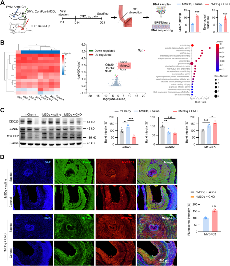
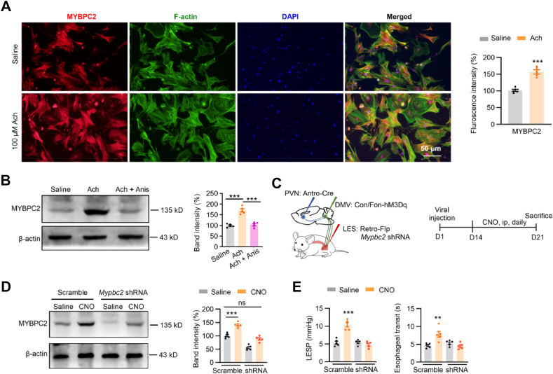
Sustained pathway enhancement changed GEJ transcription and proteins: MYBPC2 increased, while CDC20 and CCNB2 decreased. ACh also raised MYBPC2 in cultured GEJ cells, and cholinergic antagonism blocked the change. Neural communication and a local molecule were thus connected in a testable relationship.
MYBPC2 is associated with striated muscle, and GEJ samples contain mixed distal esophageal and sphincter tissue. The sampling site placed the molecular clue at the junction while leaving its exact cellular origin unresolved. Distribution across muscle components will help explain its contribution to local motility changes.
Source: Zhang K et al. · Molecular metabolism · CC BY-NC-ND
Original figure and English caption ↗
Reducing MYBPC2 improves organ outcomes after activation
Local Mybpc2 reduction improved elevated LESP and delayed transit caused by sustained pathway activation. Expression findings and functional modulation aligned, supporting MYBPC2 involvement in organ changes during this activated state. The effect of a central output could thus be followed into a peripheral molecular component.
Other proteins, inflammation and cellular components also changed within GEJ, while multiple PVN cell types and DMV outputs exist centrally. Resolving these components will clarify the brain-to-esophagus connection. The MYBPC2 findings provide a defined entry point linking neural output with organ function.
Source: Zhang K et al. · Molecular metabolism · CC BY-NC-ND
Original figure and English caption ↗
Following stress across brain, communication and organ
The study follows stress-related feeding changes into organ transit, central outputs, cholinergic signaling and peripheral molecules. The particularly clear dissociation between esophageal improvement and persistent behavioral changes shows that related states can have different regulatory sites. This provides a route from behavior into defined brain–visceral mechanisms.
Feeding is a whole-animal behavior involving finding food, initiating consumption and transporting it afterward. Examining these steps separately reveals how different mechanisms can contribute to similar reductions in feeding. Attention to organ motility adds an important layer to the relationship between stress and feeding.
The experiments mainly used adult male mice with small groups, and human and mouse esophageal muscle composition differs. Studies of cellular origin, sex and longer-term effects will extend the connection’s scope. The findings provide neural and molecular leads that can be followed across levels to understand stress-associated dysmotility.