How do blood pressure fluctuations awaken microglia in the hypothalamus?
A mouse study examines the dense microvasculature of the hypothalamic paraventricular nucleus, showing how leaked ATP activates microglia via P2Y12 receptors to drive sympathetic excitation.
Sympathetic overexcitation is a central driver of essential hypertension and end-organ damage, with central neuroinflammation widely recognized as a key contributor to autonomic dysregulation. Yet how peripheral hemodynamic disturbances in the earliest stage of hypertension are selectively sensed by specific brain nuclei across an intact blood-brain barrier and translated into sustained sympathetic outflow has remained unclear. Published in Immunity in August 2024, Peng Shi, Xiaoli Liu, Xiao Z. Shen, and colleagues at the Second Affiliated Hospital and Institute of Translational Medicine of Zhejiang University School of Medicine delineate a vascular–microglial–presympathetic neuron cascade that bridges peripheral hemodynamic stress and central sympathetic hyperactivation. Using L-NAME- and angiotensin II (Ang II)-induced mouse models of hypertension, the team shows that microglia in the hypothalamic paraventricular nucleus (PVN) undergo region-specific accumulation and pro-inflammatory activation prior to pre-sympathetic neuronal hyperexcitability. This regional vulnerability arises from the uniquely dense, ultra-small capillary network of the PVN, which facilitates parenchymal accumulation of circulating ATP under elevated hemodynamic shear stress to engage microglial P2Y12–C/EBPβ signaling and excite RVLM-projecting PVN pre-sympathetic neurons.
During early hypertension, elevated circulating ATP selectively enters the hypothalamic paraventricular nucleus (PVN) through its dense ultra-small capillary network, engaging the microglial P2Y12–C/EBPβ axis to upregulate pro-inflammatory cytokines and excite PVN–RVLM pre-sympathetic neurons ahead of systemic sympathetic hyperactivity.
Contents · 6 sections
PVN Microglial Reaction Precedes Pre-sympathetic Neuronal Hyperexcitability
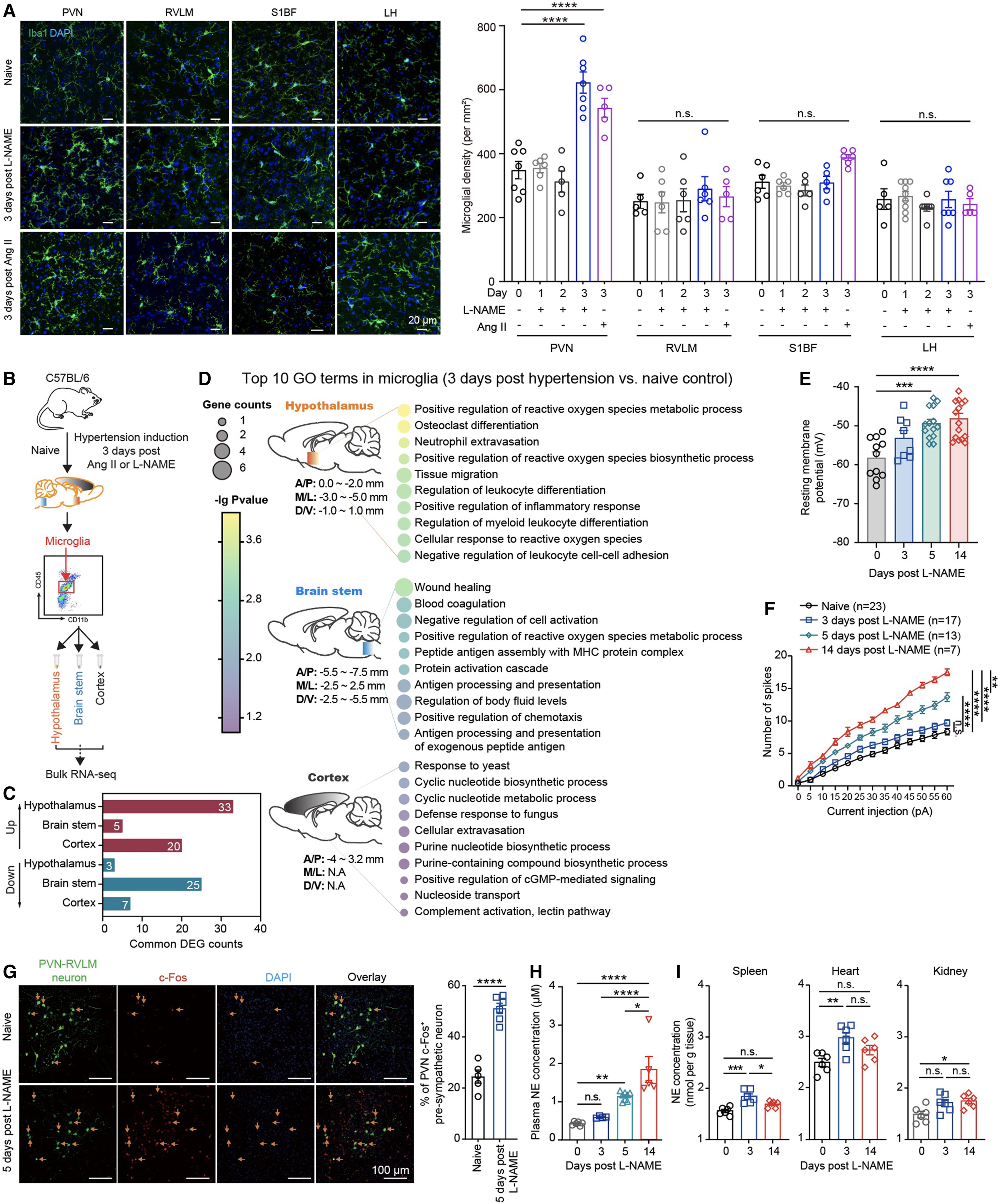
To resolve the temporal sequence between central immune activation and sympathetic excitation, the authors tracked multiple autonomic and control brain regions across two mouse models of hypertension induced by L-NAME in drinking water or subcutaneous osmotic minipump infusion of angiotensin II (Ang II). Immunofluorescence staining and 3D morphological reconstruction revealed that on day 3 of treatment, Iba1-positive microglial density increased selectively within the hypothalamic paraventricular nucleus (PVN), accompanied by enlarged soma volume and reduced process ramification. By contrast, microglial density and morphology remained unchanged at this early time point in the brainstem rostroventrolateral medulla (RVLM), lateral hypothalamus (LH), and primary somatosensory barrel cortex (S1BF).
Fluorescence-activated cell sorting coupled with low-input bulk RNA sequencing (Holo-Seq) corroborated this regional specificity: on day 3, FACS-isolated CD11b+CD45low hypothalamic microglia shared 33 commonly upregulated differentially expressed genes across both hypertension models (compared with only 5 in brainstem and 20 in cortex), enriched for reactive oxygen species metabolism, leukocyte migration, and inflammatory responses. Conversely, whole-cell patch-clamp recordings and c-Fos immunostaining of retrogradely labeled RVLM-projecting PVN pre-sympathetic (PVN–RVLM) neurons showed that resting membrane potential depolarization, elevated evoked action potential firing, and increased c-Fos expression emerged only from day 5 onward, coinciding with elevated norepinephrine (NE) levels in plasma, spleen, heart, and kidney. This clear temporal gap demonstrates that PVN microglial activation precedes systemic sympathetic hyperactivity.
Source: et al. · Immunity · Original figure reproduced for academic literature review under fair scholarly commentary; copyright remains with the authors/publisher.
Original figure and English caption ↗
Hemodynamic Disturbance Elevates Circulating ATP and Drives Selective Parenchymal Accumulation in the PVN
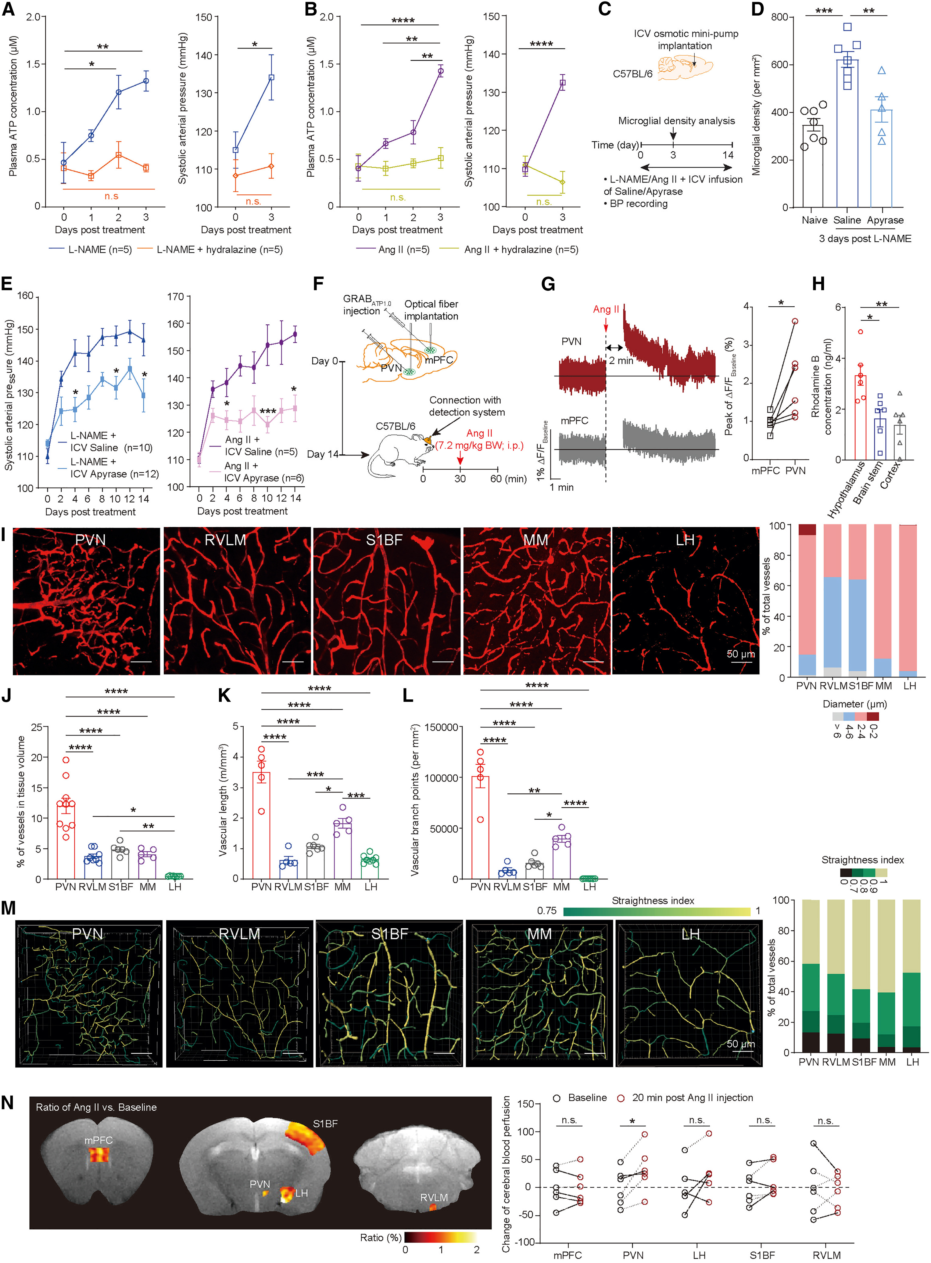
Because Ki67 and BrdU staining showed minimal local microglial proliferation on day 3 and Cx3cr1GFP/GFP mice still developed normal PVN microglial accumulation, the authors sought the upstream chemoattractant signal recruiting nearby microglia into the PVN. Vascular endothelial cells release ATP into the circulation when subjected to elevated hemodynamic shear stress. Indeed, plasma ATP levels rose significantly on days 1–2 of L-NAME or Ang II treatment, whereas co-administration of the direct vasodilator hydralazine normalized both blood pressure and plasma ATP while preventing day-3 PVN microglial accumulation. Conversely, intracerebroventricular (ICV) infusion of apyrase to hydrolyze central extracellular ATP abolished day-3 PVN microglial accumulation and restrained blood pressure elevation in both models.
To directly test in vivo whether an acute pressor challenge preferentially increases extracellular ATP in the PVN, the team expressed the genetically encoded fluorescent sensor GRAB_ATP1.0 (AAV9-CAG-cATP1.0-WPRE) simultaneously in the PVN and medial prefrontal cortex (mPFC) of the same mouse and performed dual-site fiber photometry. Within 2 minutes of an acute intraperitoneal bolus of Ang II (7.2 mg/kg), extracellular ATP fluorescence (ΔF/F) rose rapidly in both regions, with a significantly higher peak amplitude in the PVN than in the mPFC. Intravenous injection of rhodamine B (479 Da, comparable in molecular weight to ATP at 507 Da) likewise showed significantly higher parenchymal permeability in the hypothalamus than in the cortex or brainstem without leakage of 10 kDa or 70 kDa FITC-dextran, confirming selective small-molecule ATP entry across an intact blood-brain barrier.
Ultra-Small Capillary Network and Selective Hyperperfusion Underlie PVN Vulnerability
Why does circulating ATP preferentially accumulate in the PVN parenchyma? Using 150 kDa FITC-dextran vascular labeling and 3D reconstruction across the PVN, RVLM, S1BF, median mammillary nucleus (MM), and LH, the researchers discovered a distinctive microvascular architecture in the PVN. Ultra-small capillaries (< 4 μm in diameter, rich in CD13-positive pericytes and scarce in α-SMA-positive smooth muscle cells) accounted for 85.3% ± 5.6% of all vessels in the PVN, far exceeding proportions in the RVLM (33.9% ± 35.2%) and S1BF (36.1% ± 20.7%). Moreover, the PVN exhibited the largest vascular volume, longest total vessel length, highest branch-point density, and lowest vessel straightness among all examined regions.
This dense, highly branched, and tortuous capillary bed provides an expansive surface area for solute exchange and prolongs local blood transit time. Furthermore, 7.0T pseudo-continuous arterial spin labeling (pCASL) MRI demonstrated that relative cerebral blood flow (rCBF) increased selectively in the PVN 20 minutes after intraperitoneal Ang II administration, whereas rCBF in the mPFC, LH, S1BF, and RVLM remained stable. Because pericyte-covered ultra-small capillaries have limited autoregulatory buffering capacity against abrupt hemodynamic surges, selective hyperperfusion in the PVN drives both endothelial ATP production and rapid transcapillary ATP extravasation into the PVN parenchyma.
Source: et al. · Immunity · Original figure reproduced for academic literature review under fair scholarly commentary; copyright remains with the authors/publisher.
Original figure and English caption ↗
Microglial P2Y12 Deletion Prevents Pre-sympathetic Excitation and Attenuates Hypertension
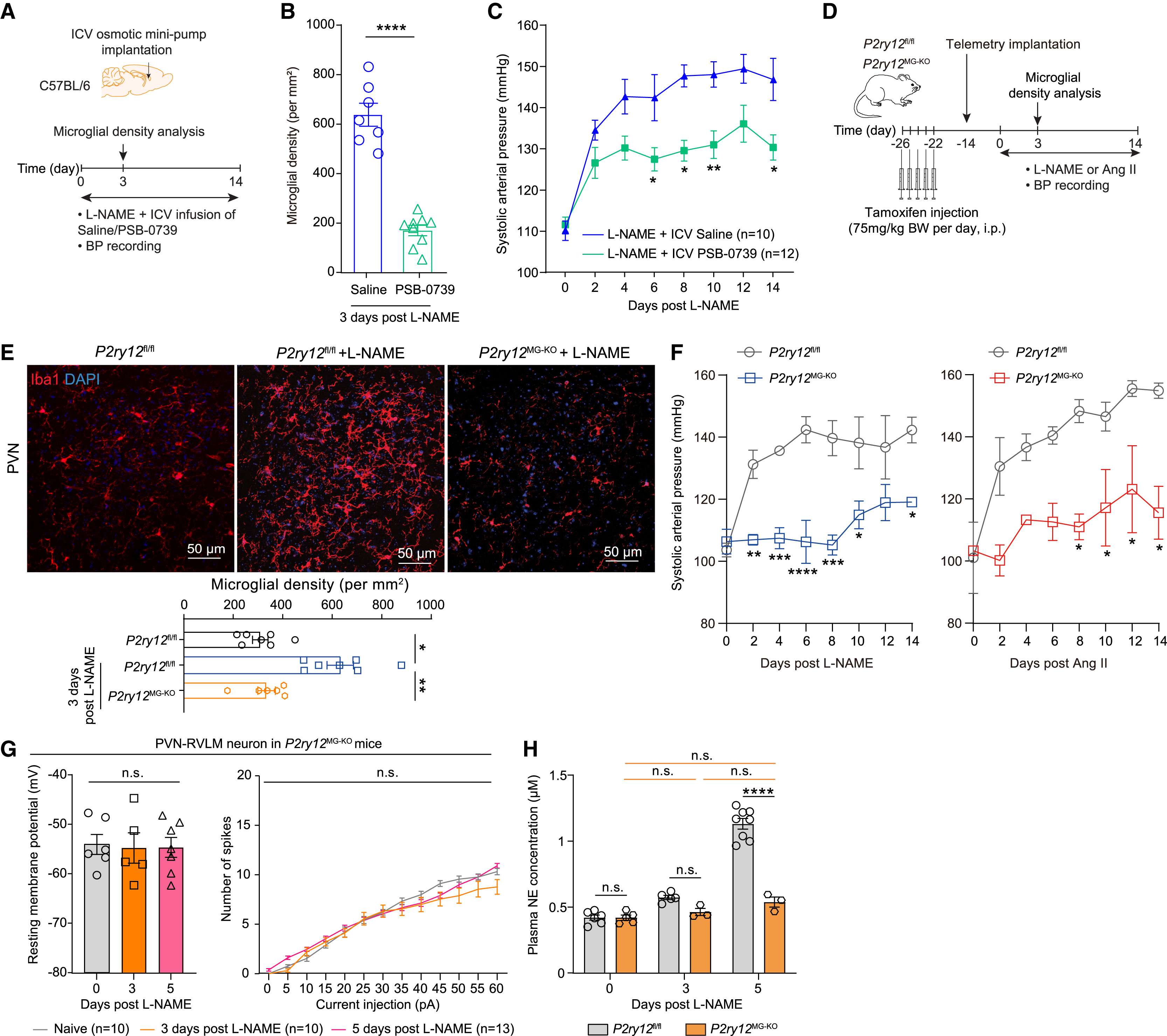
Within the central nervous system, the purinergic receptor P2Y12 is exclusively expressed on microglia and serves as the primary sensor mediating microglial chemotaxis toward extracellular ATP. To test the requirement for ATP–P2Y12 signaling in hypertension, the researchers first infused the selective P2Y12 antagonist PSB-0739 intracerebroventricularly during L-NAME administration. Pharmacological P2Y12 blockade abrogated day-3 PVN microglial accumulation and significantly blunted systolic blood pressure elevation.
To achieve genetic specificity, the team generated tamoxifen-inducible microglial P2Y12 knockout mice (Cx3cr1CreERT2/+::P2ry12fl/fl, termed P2ry12MG-KO). In both L-NAME and Ang II hypertension models, P2ry12MG-KO mice failed to develop day-3 PVN microglial accumulation and exhibited markedly attenuated blood pressure elevation compared with P2ry12fl/fl littermates. Crucially, on day 5 of L-NAME treatment, microglial P2Y12 deletion prevented resting membrane potential depolarization, high-frequency action potential firing, and c-Fos upregulation in PVN–RVLM pre-sympathetic neurons, while normalizing plasma norepinephrine levels. Conversely, infusion of the P2Y12 agonist ADPβS sufficed to increase PVN microglial density, PVN–RVLM neuronal c-Fos, and blood pressure in control mice, effects that were abolished in P2ry12MG-KO mice.
Source: et al. · Immunity · Original figure reproduced for academic literature review under fair scholarly commentary; copyright remains with the authors/publisher.
Original figure and English caption ↗
P2Y12 Activates Transcription Factor C/EBPβ to Drive Microglial Pro-inflammatory Cytokine Expression
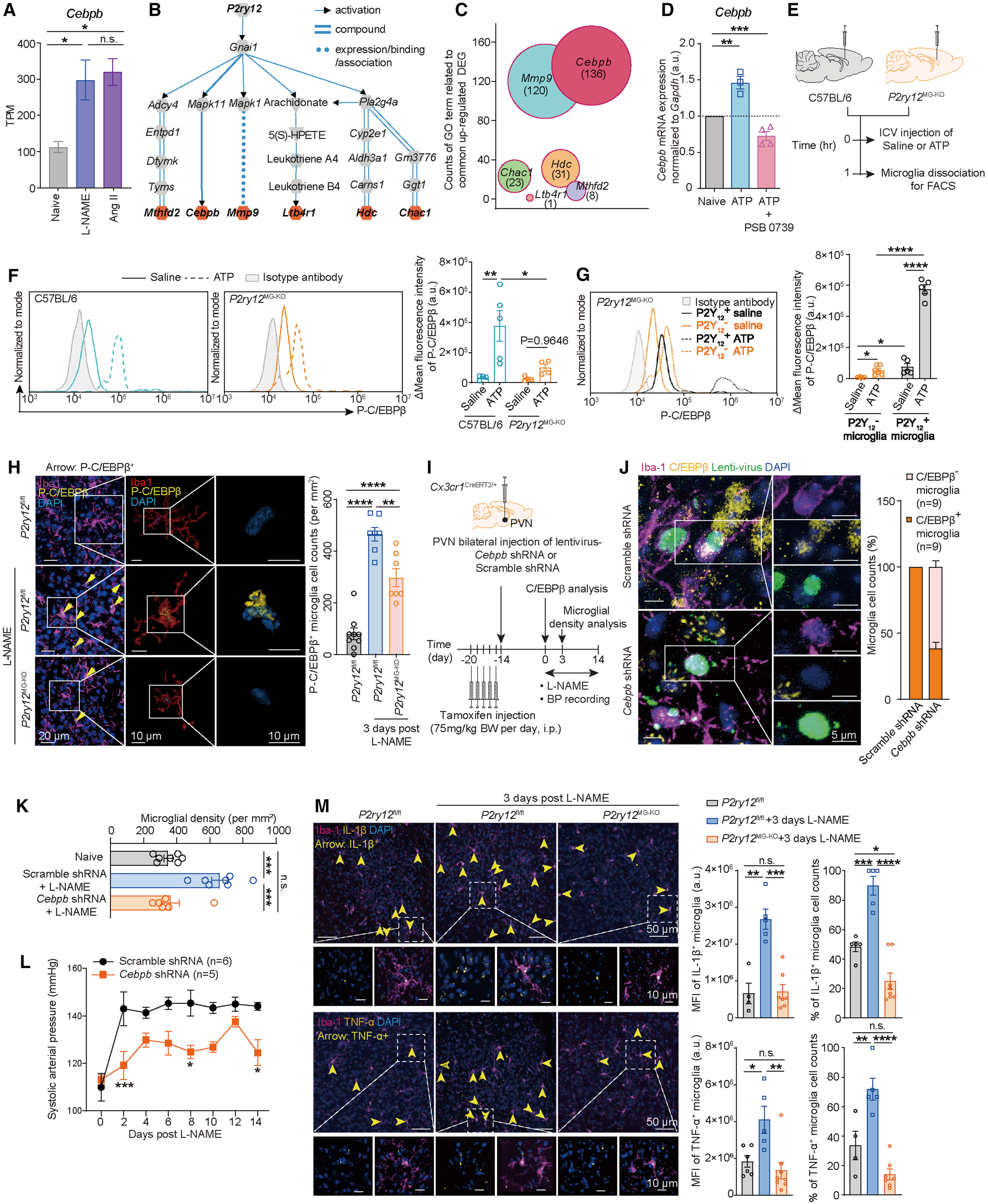
Although P2Y12 is classically viewed as a chemotactic receptor, how does its engagement trigger a pro-inflammatory transition? Intersecting the 33 commonly upregulated hypothalamic microglial genes on day 3 with KEGG downstream pathways of P2Y12 pinpointed CCAAT/enhancer-binding protein beta (C/EBPβ, encoded by Cebpb) as the sole positively regulated direct transcription factor downstream of P2Y12. Both BV2 microglial culture assays and intracerebroventricular ATP injection confirmed that ATP robustly induces Cebpb transcription and C/EBPβ Thr188 phosphorylation (P-C/EBPβ) in a P2Y12-dependent manner: 1 hour after ICV ATP injection, microglial P-C/EBPβ increased 10-fold in wild-type mice versus 2.8-fold in P2ry12MG-KO mice, and L-NAME-induced accumulation of P-C/EBPβ-positive microglia in the PVN on day 3 was similarly blunted in P2ry12MG-KO mice.
To test the causal role of microglial C/EBPβ within the PVN, the authors bilaterally injected a Cre-dependent lentivirus (rLV-CMV-DIO-shRNA(Cebpb)) into the PVN of Cx3cr1CreERT2/+ mice. Selective knockdown of C/EBPβ in PVN microglia normalized day-3 microglial density and significantly attenuated L-NAME-induced systolic blood pressure elevation. Furthermore, L-NAME treatment markedly increased the fluorescence intensity and proportion of IL-1β- and TNF-α-positive microglia in the PVN of P2ry12fl/fl mice, an effect completely abolished in P2ry12MG-KO mice. Because IL-1β and TNF-α directly potentiate excitatory drive and diminish GABAergic inhibition onto PVN pre-sympathetic neurons, these findings complete the molecular cascade linking ATP–P2Y12–C/EBPβ signaling to neuroinflammation-driven sympathetic excitation.
Source: et al. · Immunity · Original figure reproduced for academic literature review under fair scholarly commentary; copyright remains with the authors/publisher.
Original figure and English caption ↗
Cross-System Dialogue from Peripheral Hemodynamic Shear Stress to Central Sympathetic Vicious Cycle
Taken together, this study uncovers a critical positive-feedback loop in the pathogenesis of hypertension. When initial blood pressure fluctuations and hemodynamic disturbances arise, vascular endothelial cells release ATP in response to shear stress. Owing to the uniquely dense ultra-small capillary bed and selective hyperperfusion of the PVN during pressor challenges, circulating small-molecule ATP preferentially traverses the intact blood-brain barrier into the PVN parenchyma. Local and neighboring microglia sense this purinergic cue via P2Y12 receptors, migrating into the PVN and activating transcription factor C/EBPβ to produce IL-1β and TNF-α. These cytokines excite PVN–RVLM pre-sympathetic neurons, elevating systemic sympathetic tone and peripheral norepinephrine release to lock transient hemodynamic perturbations into sustained hypertension.
Methodologically, integrating dual-site in vivo fiber photometry, 3D microvascular topological quantification, and high-field arterial spin labeling MRI enabled the authors to capture the real-time transduction of acute peripheral pressor surges into nucleus-specific neurochemical signals in living mice. Beyond answering why the PVN acts as the epicenter of early neuroinflammation in hypertension, these findings highlight microvascular hemodynamic buffering and the microglial P2Y12–C/EBPβ axis as promising therapeutic targets for breaking the vicious cycle of neurogenic hypertension.