As morphine slows the small intestine, a brain circuit grows quieter
A male-mouse study traces a hypothalamus–brainstem–small-intestine connection, showing that brain opioid receptors can reduce neural activity supporting intestinal function.
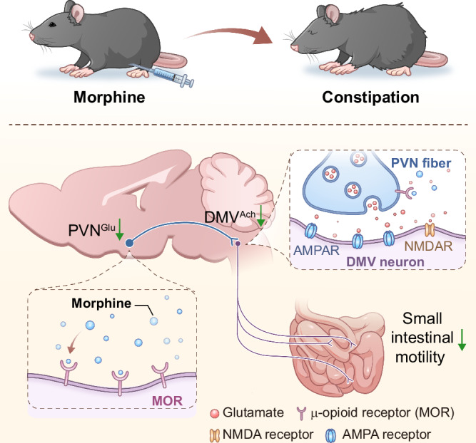
The intestine does not set its rhythm entirely on its own. Peripheral opioid receptors are a familiar explanation for morphine-associated constipation, but does the brain also contribute? A Nature Communications study in male mice identifies a route from hypothalamic paraventricular nucleus through the dorsal motor nucleus of the vagus to the small intestine. Morphine quiets this network, while raising associated activity improves intestinal function and fecal output, revealing contributions from both brain and periphery.
Morphine reduces activity through μ-opioid receptors on PVN excitatory cells, weakening associated brainstem–small-intestine drive. This central route and peripheral receptor effects both contribute to the measured constipation phenotype.
Contents · 6 sections
Slower motility does not mean weaker individual contractions
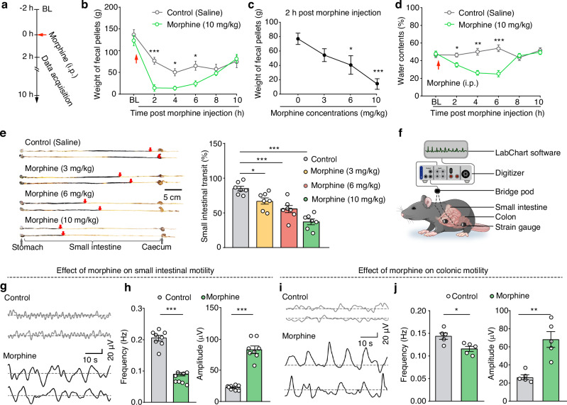
After morphine, fecal weight and water content fall, and small-intestinal contents advance more slowly. Direct motility observations show more than a uniform weakening: contraction frequency decreases while recorded amplitude increases. Reduced fecal output accompanies a less favorable pattern for propulsion.
The colon also shows lower frequency and greater amplitude, while total food and water intake remain without significant changes. The study follows the neural connections regulating the gut. The vagus is an important brain–digestive tract channel, with the dorsal motor nucleus of the vagus (DMV) serving as a key descending node.
Source: Ma J et al. · Nature communications · CC BY
Original figure and English caption ↗
Brainstem cells linked to the small intestine become less active
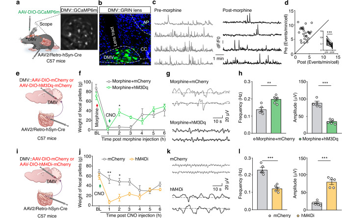
Anatomical observations connect DMV cholinergic cells with the small intestine, where fibers from this population are visible. Raising the activity of associated intestinal terminals increases motility frequency and lowers amplitude under morphine. The connection in tissue images thus gains a functional organ response.
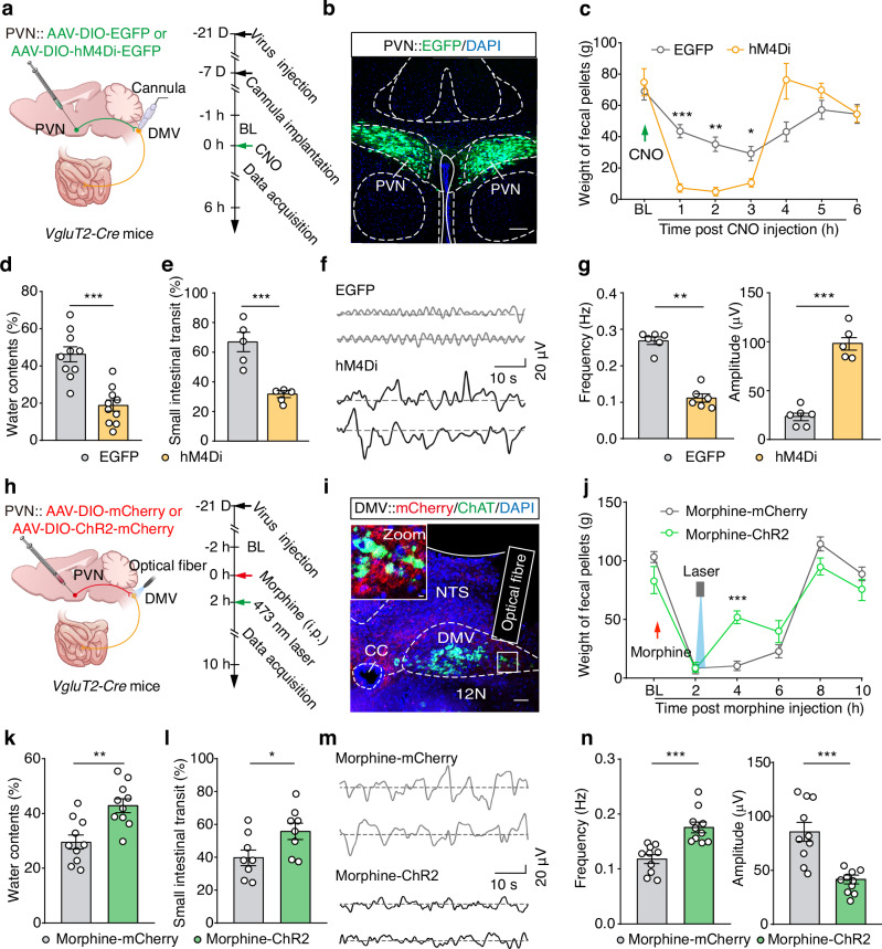
Morphine reduces firing and calcium activity in DMV cells linked to the small intestine. Activating the population improves fecal weight, water content and transit; inhibiting it produces a similar constipation phenotype in mice not given morphine. Brainstem activity therefore participates in regulation rather than simply accompanying organ changes.
This node links brain activity with the rhythm of intestinal propulsion. Fecal output is not a simple copy of one contraction recording: transit, water content and motility patterns jointly shape it. Convergent changes across readouts clarify the functional contribution of the brainstem population.
Source: Ma J et al. · Nature communications · CC BY
Original figure and English caption ↗
The hypothalamus provides excitatory brainstem input
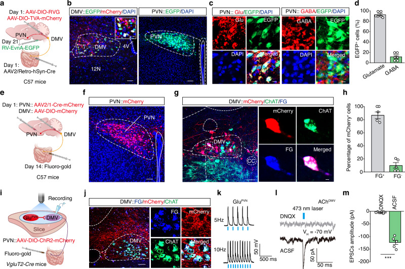
Tracing upstream identifies several regions, with substantial labeling in paraventricular nucleus (PVN), where associated cells are predominantly excitatory. Complementary anatomical observations connect PVN, DMV cholinergic cells and small intestine, bringing nodes at different levels into one testable route.
Physiological observations show that PVN input evokes excitatory synaptic currents in small-intestine-linked DMV cells. A sustained NMDA-receptor-associated current decreases after morphine and also after reducing PVN excitatory cells. These findings support an upstream contribution to ongoing brainstem drive.
Source: Ma J et al. · Nature communications · CC BY
Original figure and English caption ↗
How does morphine quiet this route?
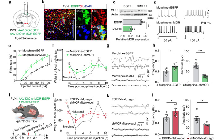
Associated PVN cells show μ-opioid receptor (MOR) labeling. Morphine lowers calcium activity in the DMV-projecting PVN population, while electrophysiology also shows reduced excitatory-cell activity. Changing opioid-receptor effects locally in PVN alters fecal output and small-intestinal motility, indicating a functional central receptor contribution.
Reducing MOR in PVN excitatory cells preserves higher firing under morphine and improves small-intestinal transit, motility and fecal measures. Colonic motility does not show the same significant improvement. Different digestive segments can therefore be neurally regulated without sharing an identical upstream control mechanism.
Receptors in different locations can change different parts of the response to the same drug. Reducing receptor expression in the relevant brain cells changes organ readouts, connecting receptor labeling with functional contribution.
Source: Ma J et al. · Nature communications · CC BY
Original figure and English caption ↗
Brain and periphery can shape the same outcome
Peripheral receptors also matter. Peripheral opioid-receptor blockade partially improves constipation; reducing associated PVN MOR alongside it further improves some measures. The central route and peripheral receptor effects therefore represent two contributions to the same phenomenon.
Increasing activity along the PVN–DMV connection improves fecal water content, small-intestinal transit and motility after morphine, while inhibition lowers these measures. Upstream drive reaches organ function through brainstem and descending connections, giving the broad brain–gut concept a specific network contribution.
Source: Ma J et al. · Nature communications · CC BY
Original figure and English caption ↗
Small-intestinal function carries a trace of brain state
Studying morphine responses in young male mice, the work links receptors, cellular activity and organ function. Its new insight is that a drug’s intestinal effects can involve reduced brain activity supporting descending output, with peripheral and central effects contributing to one outcome.
The small-intestine–colon distinction is especially revealing. Slower gut function can involve different segments and regulatory routes, with fecal output emerging where those changes converge. Placing an organ within its neural network extends the mechanistic picture from local receptors to connections spanning brainstem and hypothalamus.