A more local view of neural cells: AAV13 as a research-tool option
In the tested regions and conditions in male mice, AAV13 labeling was more spatially confined than AAV2. The study also demonstrated localized sparse labeling and behavior-associated calcium recording.
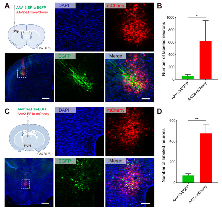
In densely packed brain tissue, labeling more cells can make a local question harder to answer. A small region may be difficult to distinguish from its neighbors when labeling extends beyond its boundaries. A study published in International Journal of Molecular Sciences on October 24, 2022 examined AAV13 as a neural research vector. In male mice, it showed relatively confined labeling in several regions and demonstrated sparse-cell observation and behavior-associated activity recording. The contribution is a research-tool option for localized observation.
AAV13 showed localized labeling in the tested conditions and supported sparse morphological observation and calcium recording. Spatial confinement and signal strength are distinct properties that require question-specific evaluation.
Contents · 6 sections
Small regions make observational boundaries important
Many neuroscience questions require distinguishing nearby populations with different functions. A clear fluorescence image does not by itself identify every contributing cell. Where labeling extends and which cells are included affect interpretation, especially in small nuclei.
A research vector cannot be evaluated solely by brightness or cell count. Broad coverage can help population studies, while confined coverage can suit local questions. This work emphasizes spatial extent in specific regions rather than establishing one best option for every experiment.
More confined labeling in the tested regions
The study compared AAV13- and AAV2-associated labeling in primary somatosensory cortex and the paraventricular hypothalamic nucleus. AAV13-associated green labeling appeared more confined, while AAV2-associated red labeling extended more broadly. AAV13 also labeled fewer positive cells in both regional comparisons.
These findings support localized labeling in the tested conditions. Fewer labeled cells do not inherently mean better performance, and cell count is not spatial volume. Different fluorescent reporters were used, and the authors noted lower AAV13 signal intensity. Confinement and brightness therefore need to be considered separately.
Source: Han Z et al. · International journal of molecular sciences · CC BY
Original figure and English caption ↗
Sparse labeling makes room for morphology
The study also demonstrated localized sparse labeling. Dense fluorescence from overlapping cell bodies and processes can make it difficult to assign a fine structure to its source. Clearly showing fewer cells serves morphological observation, a different goal from broad population coverage.
Images across genetic backgrounds and labeling conditions show that the capability was not limited to one representative field. Visible processes do not, however, establish a complete whole-brain reconstruction of an individual neuron. Full trajectories and connectivity require additional evidence.
Predominantly neuronal does not mean absolutely exclusive
In tested regions, most positive labeling aligned with neuronal markers, while a small amount of astrocyte-associated labeling was also observed. Cellular composition matters alongside spatial distribution: a localized field does not guarantee that only one cell class contributes.
Locality, sparseness and cell-class preference are related but distinct properties. The work provides initial observations across these dimensions without establishing specificity across every brain region and cellular state. Interpretation must remain consistent with the actual labeled population.
Recording a population signal during behavior
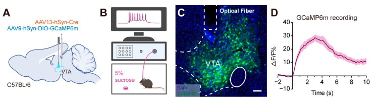
The team also demonstrated calcium recording in the ventral tegmental area. Population calcium signals rose around sucrose licking, providing an application example for behavior-associated functional observation rather than static tissue imaging alone.
A calcium increase around behavior does not establish that these cells cause reward experience. Nor can a population signal automatically be assigned to each cell. The VTA contains diverse populations, so its location does not identify every recorded cell as dopaminergic.
Source: Han Z et al. · International journal of molecular sciences · CC BY
Original figure and English caption ↗
Another option with a defined scope
The contribution is to evaluate localized observation, sparse morphology and functional recording within one research-tool study. It establishes neither a human disease treatment nor superiority in every task. The findings concern adult male mice, tested regions and particular reporter conditions.
Why labeling is more confined, and how signal strength relates to spatial extent, remain mechanistic questions. Within those limits, a different spatial profile can still be useful when a research question calls for observing a small local population.