When a trauma cue returns, how does ketamine weaken social avoidance traces in the amygdala?
Focusing on the memory reconsolidation window in mice, researchers show that subanesthetic esketamine recruits midbrain dopamine input and amygdala D2 receptors to attenuate trauma engrams.
Patients with post-traumatic stress disorder (PTSD) frequently experience severe relapse of fear and social avoidance upon encountering trauma-related reminders, even long after initial symptoms appear to have subsided. Selectively erasing stored traumatic memory engrams to prevent long-term relapse remains a major challenge in neuroscience and psychiatry. Published in Neuron in July 2024, Bo Tian, Pei Zhang, Jie Ming, and colleagues at Tongji Medical College of Huazhong University of Science and Technology established a mouse model of acute social defeat stress (SDS) followed by 4 weeks of solitary housing that recapitulates short-term social avoidance, natural forgetting, and cue re-exposure-induced long-term relapse. The authors demonstrate that traumatic social memories are encoded in engram cells within the basolateral amygdala (BLA), and that a single subanesthetic dose of (S)-ketamine (5 mg/kg) administered immediately after trauma cue re-exposure—within the memory reconsolidation window—activates monosynaptic ventral tegmental area (VTA) dopaminergic inputs onto BLA engram cells via D2 receptors (D2Rs) to silence engram reactivation and prevent both short- and long-term social avoidance.
Administered immediately within the memory reconsolidation window following trauma cue re-exposure, a single subanesthetic dose of (S)-ketamine (5 mg/kg) activates monosynaptic VTA dopaminergic projections onto BLA traumatic engram cells and engages local D2 receptors to remodel synaptic plasticity pathways, silence engram reactivation, and abolish short- and long-term social avoidance.
Contents · 5 sections
Naturally Forgotten Traumatic Social Memory Persists in Latent BLA Engram Cells
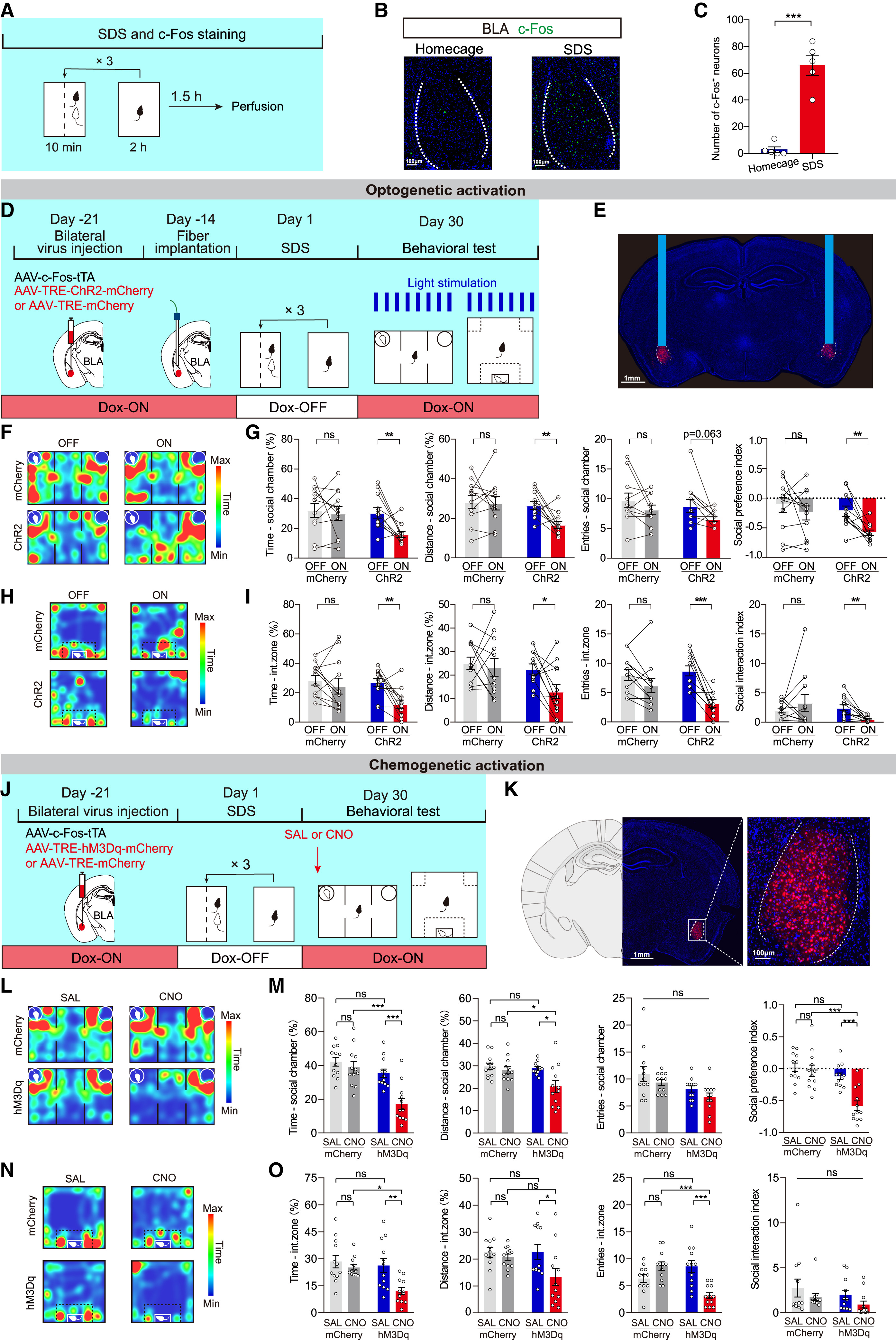
To distinguish superficial behavioral remission from true erasure of a traumatic memory trace, the authors established an acute social defeat stress (SDS) paradigm combined with 4 weeks of solitary housing. Male C57BL/6J mice subjected to three 10-minute bouts of social defeat by aggressive CD-1 mice on Day 1 exhibited robust short-term social avoidance on Day 2 in both the three-chamber social interaction test and open-field social preference-avoidance test. After 4 weeks of solitary housing, this avoidance naturally dissipated by Day 30. However, a 10-minute vicarious trauma re-exposure on Day 31—watching a conspecific C57 mouse being defeated by a CD-1 aggressor across a perforated divider—potently reinstated long-term social avoidance, whereas re-exposure alone without prior SDS had no effect.
Because acute SDS strongly induced c-Fos expression in the basolateral amygdala (BLA), the researchers used an activity-dependent Tet-Off tagging system (AAV9-c-Fos-tTA plus AAV9-TRE-ChR2-mCherry or AAV9-TRE-hM3Dq-mCherry under doxycycline withdrawal during Day 1 SDS) to capture SDS-activated BLA engram cells. On Day 30, when behavioral avoidance had naturally subsided, either optogenetic (20 Hz blue light) or chemogenetic (CNO) reactivation of these SDS-tagged BLA engram cells was sufficient to reinstate severe social avoidance without affecting general sociability toward C57 strangers, whereas activating randomly labeled BLA neurons had no effect. Thus, natural forgetting leaves the latent traumatic memory trace intact within BLA engram cells.
Source: et al. · Neuron · Original figure reproduced for academic literature review under fair scholarly commentary; copyright remains with the authors/publisher.
Original figure and English caption ↗
Subanesthetic (S)-Ketamine Administered Immediately After Cue Re-exposure Erases Traumatic Avoidance
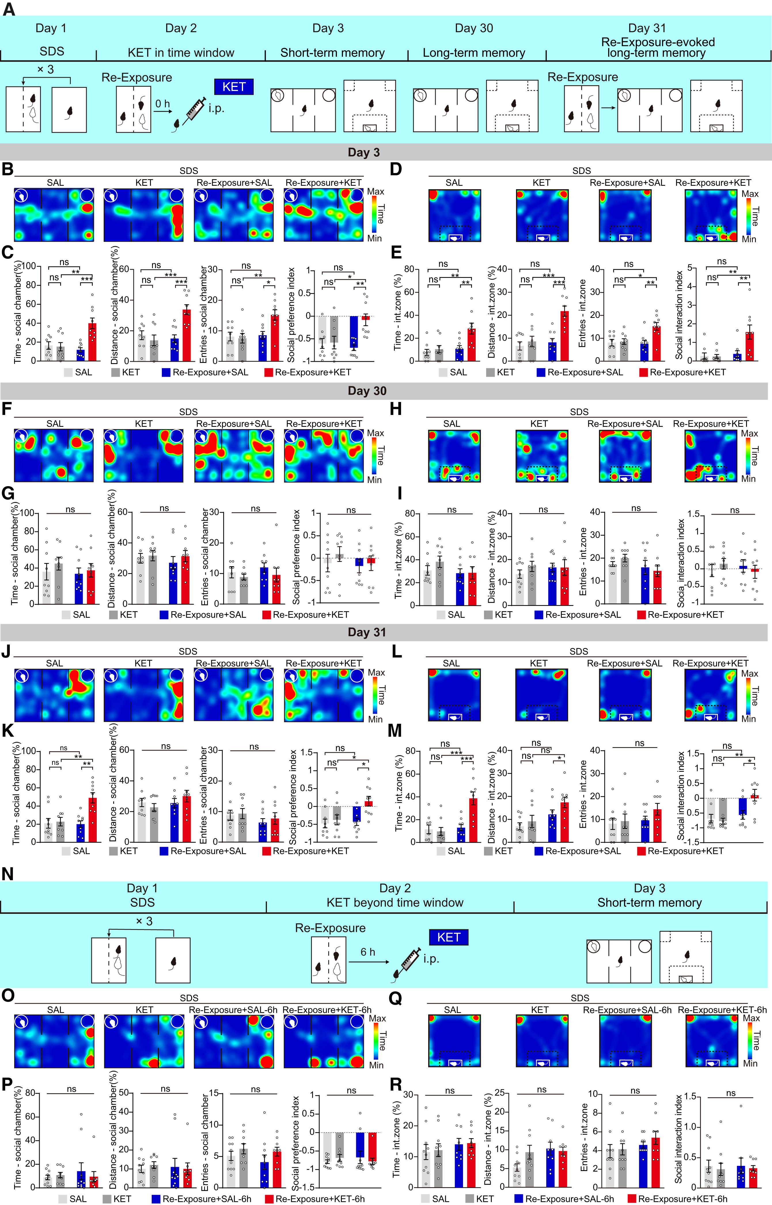
According to memory reconsolidation theory, retrieving a consolidated memory renders it transiently labile for several hours, creating a window during which pharmacological intervention can disrupt reconsolidation and erase the memory trace. Testing a combined 'Re-Exposure + KET' paradigm on Day 2 after SDS, the authors exposed mice to 10 minutes of vicarious trauma re-exposure followed immediately (0 hours) by a single intraperitoneal injection of subanesthetic (S)-ketamine (5 mg/kg). Matching the positive control rapamycin, Re-Exposure + KET markedly alleviated short-term social avoidance on Day 3 and completely prevented cue-induced relapse of long-term social avoidance on Day 31.
Control conditions underscored the strict requirement for memory retrieval and timing: neither (S)-ketamine alone without re-exposure (KET) nor re-exposure followed by saline (Re-Exposure + SAL) alleviated social avoidance on Day 3 or prevented relapse on Day 31. Crucially, delaying (S)-ketamine administration until 6 hours after re-exposure—outside the memory reconsolidation window—completely abolished its therapeutic effect. Because immediate post-retrieval (S)-ketamine also accelerated extinction in an auditory fear conditioning paradigm, these results establish that subanesthetic (S)-ketamine acts specifically by disrupting traumatic memory reconsolidation.
Source: et al. · Neuron · Original figure reproduced for academic literature review under fair scholarly commentary; copyright remains with the authors/publisher.
Original figure and English caption ↗
In Vivo Fiber Photometry Reveals an 80% Reduction in BLA Engram Calcium Reactivity After Re-exposure Plus Ketamine
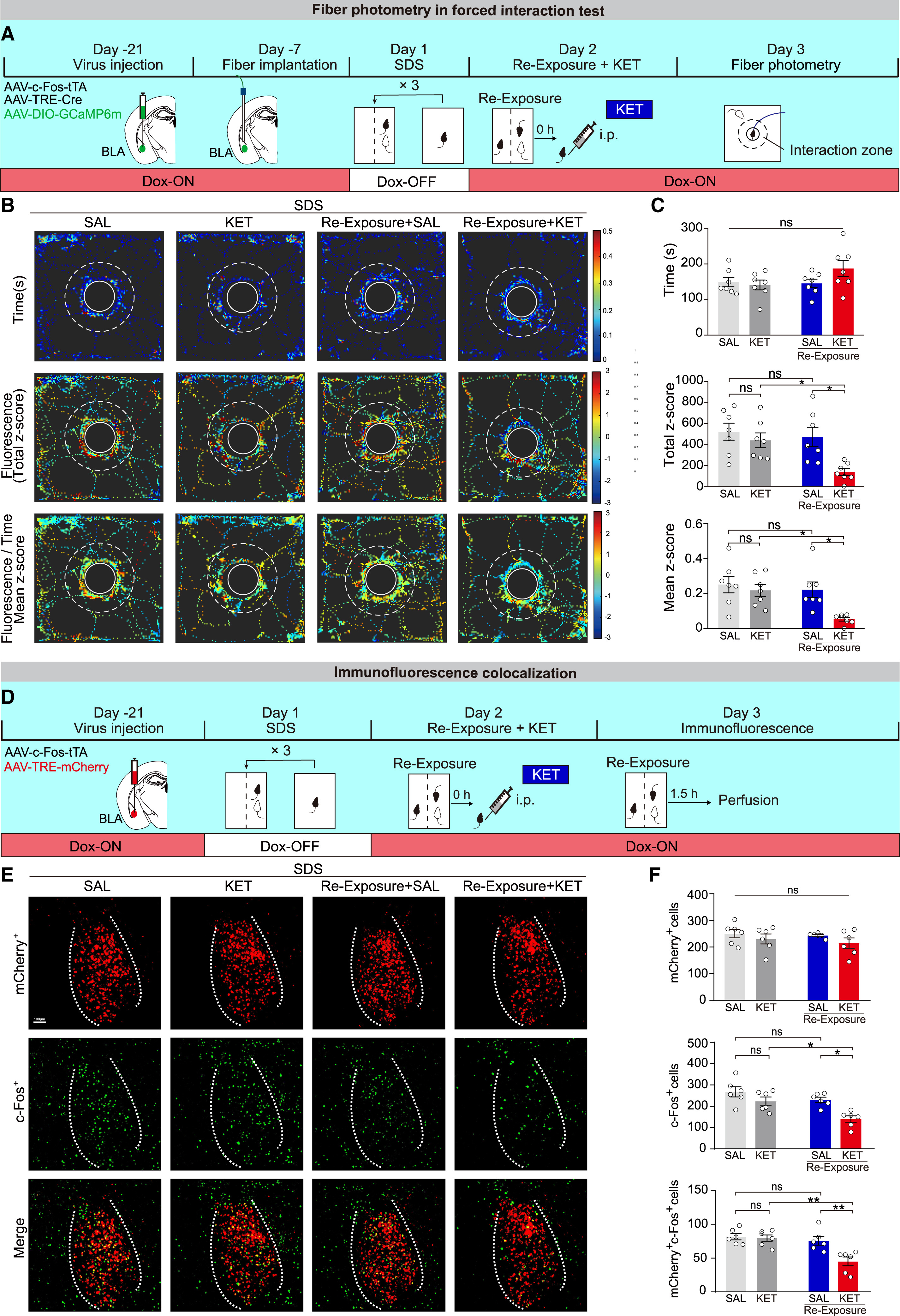
How does Re-Exposure + KET alter the functional reactivity of BLA traumatic engram cells in vivo? Using Tet-Off activity-dependent labeling (AAV9-c-Fos-tTA + AAV9-TRE-Cre + AAV9-DIO-GCaMP6m) to express GCaMP6m selectively in Day 1 SDS-activated BLA engram cells, the researchers performed in vivo fiber photometry (473 nm signal and 405 nm isosbestic control) during a forced interaction test with a freely moving CD-1 mouse on Day 3. Although interaction duration with the CD-1 mouse was comparable across groups, only the Re-Exposure + KET group exhibited a dramatic (~80%) reduction in both total and time-adjusted mean GCaMP6m calcium fluorescence (Z-score) in BLA engram cells upon CD-1 entry into the interaction zone, whereas randomly labeled BLA neurons showed no calcium response.
Double immunofluorescence staining corroborated these real-time calcium dynamics at the cellular population level. Tagging Day 1 SDS engram cells with mCherry (AAV9-c-Fos-tTA + AAV9-TRE-mCherry) and staining for endogenous c-Fos 1.5 hours after Day 3 re-exposure revealed that while total mCherry-positive engram cell counts remained unchanged across groups, the number of reactivated engram cells (mCherry+c-Fos+) and total c-Fos+ cells dropped by approximately 50% selectively in the Re-Exposure + KET group. Together, fiber photometry and histological mapping demonstrate that Re-Exposure + KET functionally silences BLA traumatic engram cells.
Source: et al. · Neuron · Original figure reproduced for academic literature review under fair scholarly commentary; copyright remains with the authors/publisher.
Original figure and English caption ↗
VTA Dopaminergic Neurons Form Functional Monosynaptic Projections onto BLA Engram Cells
Through what upstream circuit does systemic (S)-ketamine remodel BLA engram cells? In vivo multi-channel electrophysiological recordings showed that intraperitoneal (S)-ketamine (unlike rapamycin) significantly increased the firing rate of putative VTA dopaminergic (VTADA) neurons. To test whether VTADA neurons directly innervate BLA traumatic engram cells, the authors used SDS-tagged BLA engram cells as starter cells for rabies-based trans-monosynaptic retrograde tracing (AAV9-c-Fos-tTA + AAV9-TRE-Cre + AAV9-DIO-TVA-EGFP + AAV9-DIO-RVG followed by RV-EnvA-ΔG-dsRed). Whole-brain mapping and tyrosine hydroxylase (TH) immunostaining confirmed direct monosynaptic inputs from TH-positive VTA dopaminergic neurons onto BLA engram cells, complemented by dense axonal terminal labeling in the BLA following anterograde AAV9-DIO-EGFP tracing in DAT-Cre mice.
To functionally validate dopamine release along this VTADA→BLA pathway in vivo, the team expressed ChR2-mCherry (AAV9-DIO-ChR2-mCherry) in VTA dopaminergic neurons of DAT-Cre mice and co-expressed the genetically encoded dopamine sensor DA3m (AAV9-Syn-Cre + AAV9-DIO-DA3m) in the ipsilateral BLA. Combining optogenetic stimulation (473 nm, 20 Hz) of VTADA neurons with in vivo fiber photometry in the BLA demonstrated robust, time-locked surges in DA3m fluorescence (ΔF/F Z-score) relative to the 405 nm isosbestic control channel, confirming functional dopamine release in the BLA.
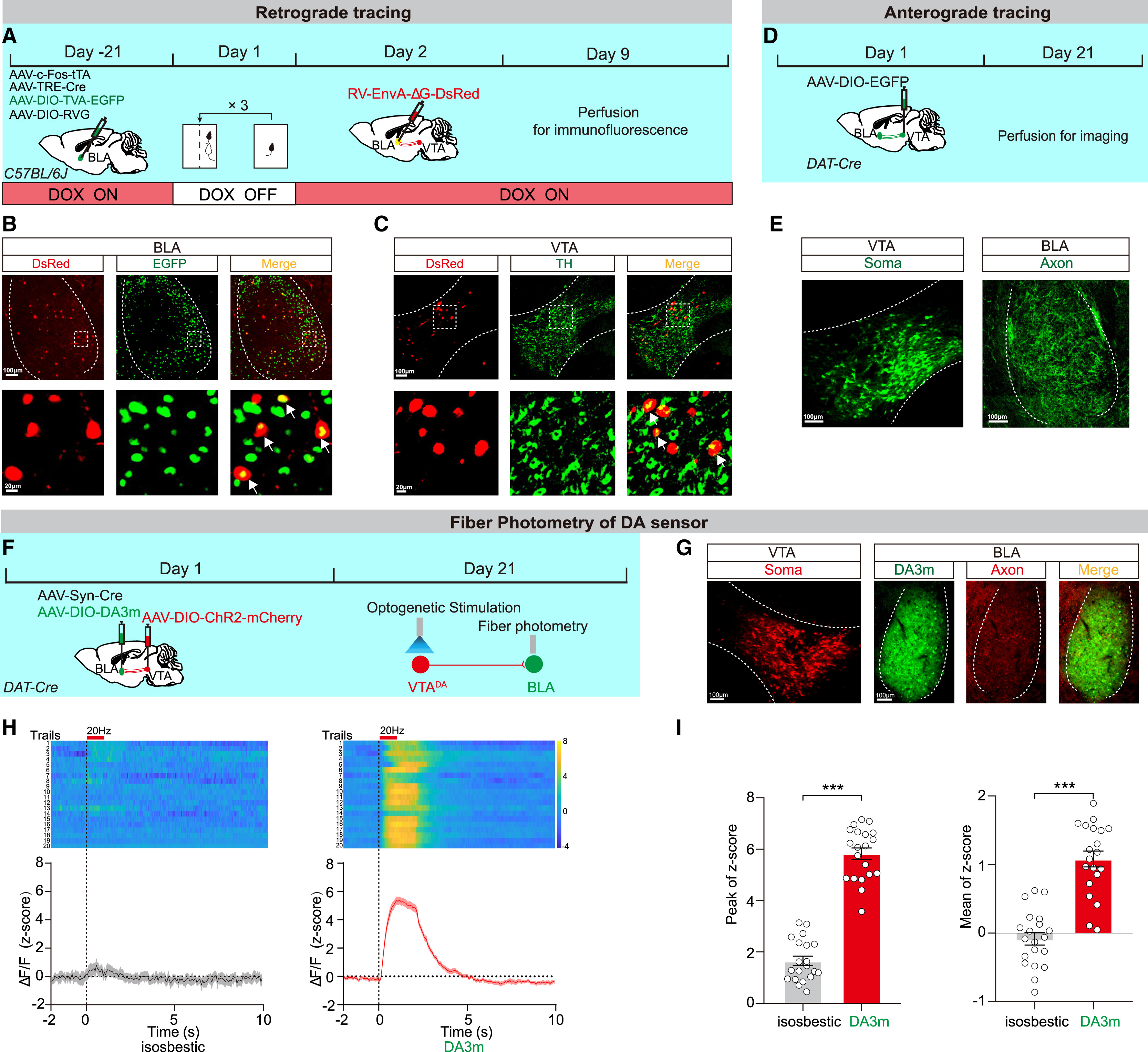
Source: et al. · Neuron · Original figure reproduced for academic literature review under fair scholarly commentary; copyright remains with the authors/publisher.
Original figure and English caption ↗
The VTADA→BLA Circuit Engages D2 Receptors and Remodels Synaptic Plasticity Transcriptomes to Erase Traumatic Memory
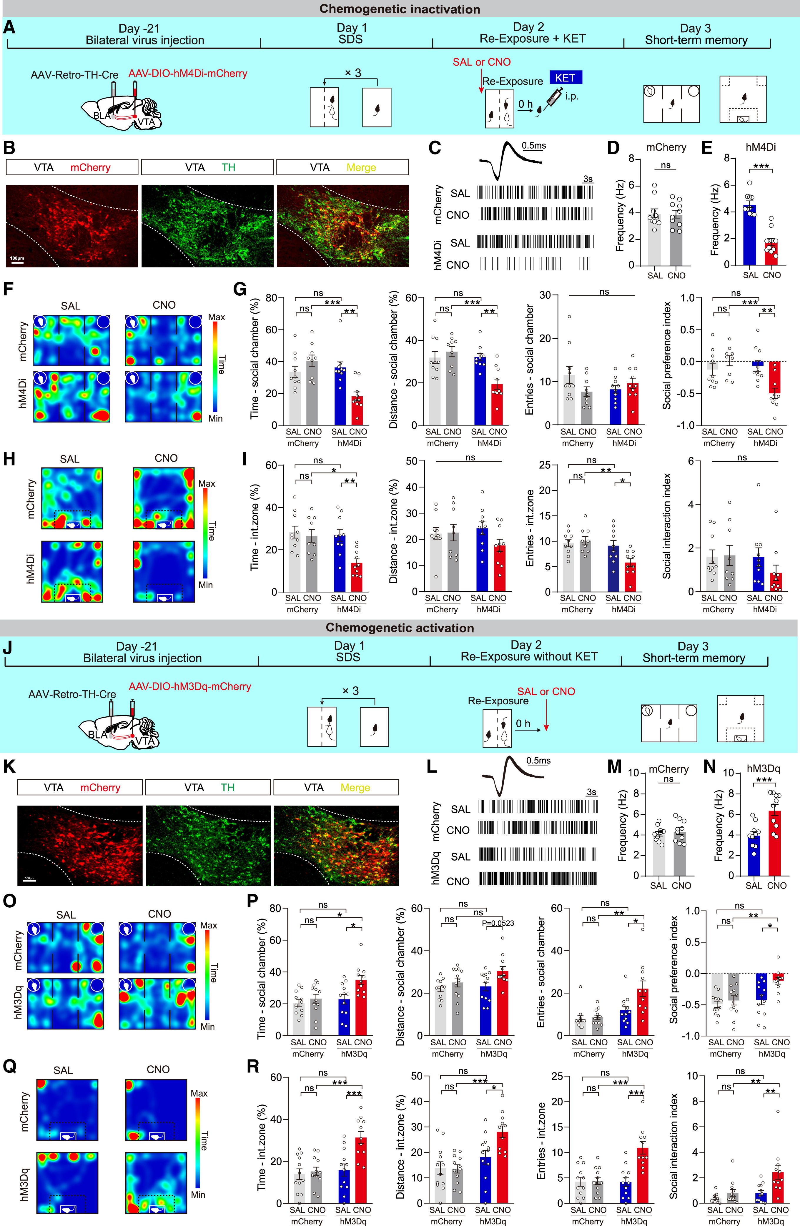
Projection-specific chemogenetic manipulations established both the necessity and sufficiency of the VTADA→BLA pathway. Injecting AAV2-Retro-TH-Cre into the BLA and AAV9-DIO-hM4Di-mCherry into the VTA to inhibit BLA-projecting VTADA neurons prior to Re-Exposure + KET completely blocked the therapeutic alleviation of both short-term (Day 3) and long-term (Day 31) social avoidance. Conversely, chemogenetic activation (AAV9-DIO-hM3Dq-mCherry + CNO) of BLA-projecting VTADA neurons immediately after trauma re-exposure—without ketamine—mimicked Re-Exposure + KET by abolishing both short- and long-term social avoidance. Furthermore, bilateral intra-BLA microinfusion of the dopamine D2 receptor (D2R) antagonist sulpiride (2 μg/side), but not the D1R antagonist SCH-23390 (1.5 μg/side), abolished both short- and long-term therapeutic effects of Re-Exposure + KET.
Finally, 10x Genomics single-nucleus RNA sequencing (snRNA-seq) of 47,997 BLA nuclei from Re-Exposure + KET versus SDS control mice identified 5 non-neuronal clusters and 10 neuronal subtypes (Penk, Crim1, Nxph1, Foxp2, Tac1, Rmst, Sst, Vip, Rorb, and Lhx8). KEGG enrichment analysis revealed 27 shared pathways across all 10 neuronal clusters and significant remodeling of memory- and synapse-related pathways across 9 of the 10 neuronal subtypes (all except Crim1)—including glutamatergic synapse, long-term depression (LTD), cAMP signaling, retrograde endocannabinoid signaling, calcium/MAPK/PI3K-Akt signaling, cell adhesion molecules, and regulation of the actin cytoskeleton. These single-cell transcriptomic findings show how D2R-mediated dopaminergic signaling rewires synaptic plasticity programs in the BLA to destabilize and erase traumatic memory engrams.