Two inputs reach the amygdala but differ in fear learning
During mouse fear learning, LPB input to the amygdala contributes during acquisition, whereas PVT input yields different intervention results. CB1 receptors also regulate excitatory transmission from LPB.
An initially harmless sound can make mice freeze after pairing with an aversive stimulus. Learning links sensory information with experience in the brain. A 2024 iScience study compares two inputs to the central lateral amygdala, CeL: lateral parabrachial nucleus, LPB, and paraventricular thalamus, PVT. Both reach CeL and show increased activity labeling after fear conditioning, yet their intervention results differ. Following recipient cells and CB1 receptors reveals circuit distinctions and synaptic regulation in fear learning.
Suppressing LPB-to-CeL terminals during learning weakens later contextual and cued fear responses; corresponding PVT-terminal suppression has no significant effect. CB1 activation reduces LPB-evoked excitatory currents, while LPB receptor knockdown does not significantly counter local pharmacological behavioral effects.
Contents · 7 sections
Context and sound become distinct cues
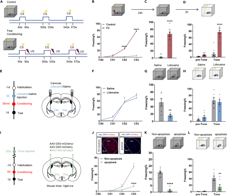
After conditioning, freezing increases in the stimulus-associated context and during the paired sound. Temporarily suppressing overall CeL activity during learning weakens subsequent contextual responses, while cued responses and training freezing change nonsignificantly.
Reducing CeL GABA cells lowers freezing at the end of training and in both subsequent tests. Transient regional suppression and cell-population reduction yield different outcomes, motivating more precise identification of populations: intervention targets and duration matter even within one region.
Source: Gao JH et al. · iScience · CC BY-NC-ND
Original figure and English caption ↗
SOM-marked cells become more responsive
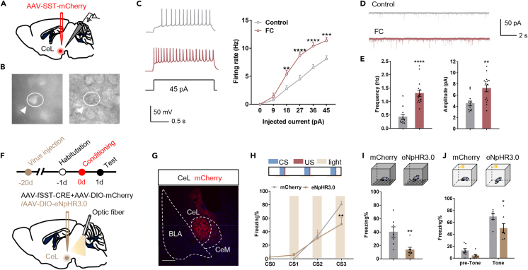
The study focuses on somatostatin-positive CeL cells, the SOM population. After conditioning, firing responses to injected current increase, as do the frequency and amplitude of received miniature excitatory currents. Both cellular responsiveness and incoming transmission change after experience.
Suppressing SOM cells during learning reduces freezing during the final training cue and weakens subsequent contextual and cued responses. Their contribution to the tested learning process prompts a search for upstream excitatory inputs.
Source: Gao JH et al. · iScience · CC BY-NC-ND
Original figure and English caption ↗
Both reach CeL among a broader set of inputs
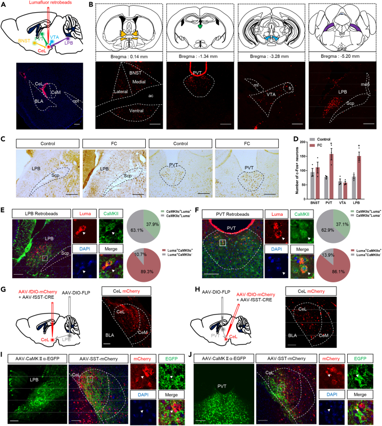
Retrograde tracing identifies several CeL input sources, including LPB, PVT, bed nucleus of the stria terminalis and ventral tegmental area. Activity labeling rises in LPB and PVT after conditioning, while corresponding changes in the other two regions are nonsignificant, focusing attention on the two excitatory inputs.
Most labeled projection cells in the two sources carry an excitatory marker, and their axons lie close to CeL SOM cells. Connectivity and increased activity are starting observations; interventions test which inputs contribute during learning.
Source: Gao JH et al. · iScience · CC BY-NC-ND
Original figure and English caption ↗
Terminal suppression during learning weakens later responses
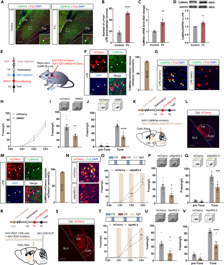
Suppressing CeL-projecting LPB cells reduces later contextual and cued responses without significantly changing training freezing. Briefly suppressing LPB terminals in CeL weakens freezing later in training and in both subsequent tests. The two approaches support a contribution to learning.
Suppressing LPB-input-tagged CeL SOM cells also weakens subsequent responses. Tested mechanical thresholds and Y-maze performance change nonsignificantly. The findings link LPB input with a recipient population and distinguish its effects from those measured sensory and spatial-behavioral outcomes.
Source: Gao JH et al. · iScience · CC BY-NC-ND
Original figure and English caption ↗
Different intervention sites and durations yield different outcomes
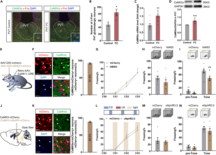
Longer-lasting suppression of CeL-projecting PVT cells weakens later cued responses, while contextual and training outcomes change nonsignificantly. Suppressing PVT terminals in CeL only during learning has no significant effect in training, contextual or cued tests. It does not reproduce the LPB-terminal result.
The PVT interventions differ in location and duration, and targeting projection cells can involve additional outputs. The authors propose a contribution to post-learning processing, requiring direct tests of post-learning time windows. The current findings show that the contributions of the two inputs are not interchangeable.
Source: Gao JH et al. · iScience · CC BY-NC-ND
Original figure and English caption ↗
CB1 receptors occur near both inputs
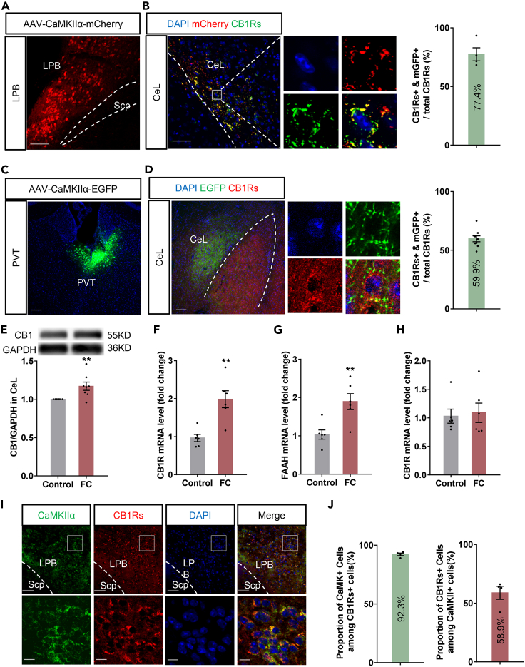
CB1-receptor labeling occurs near both LPB and PVT terminals in CeL. After conditioning, CeL CB1 protein and LPB receptor transcripts rise, while PVT transcripts change nonsignificantly. This molecular difference accompanies the distinct behavioral results of the two inputs.
CB1 participates in endocannabinoid regulation and can alter synaptic transmission. Increased expression does not itself establish stronger receptor signaling, so the study stimulates LPB input in slices to observe recipient currents directly.
Source: Gao JH et al. · iScience · CC BY-NC-ND
Original figure and English caption ↗
Input regulation extends beyond one pathway
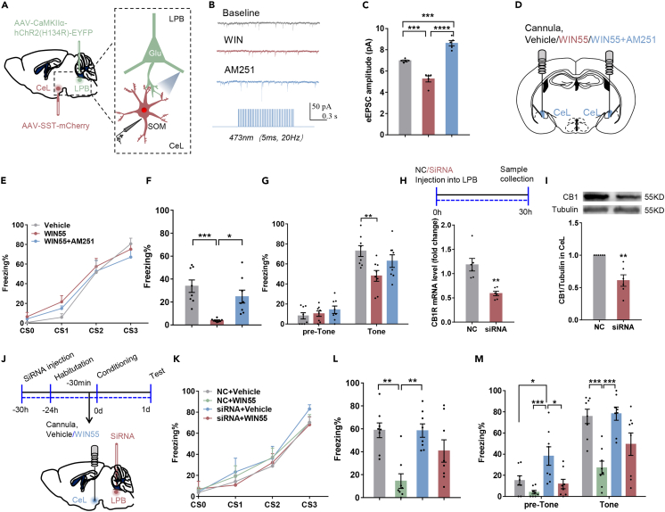
CB1-related activation reduces SOM-cell excitatory currents evoked by LPB stimulation, whereas receptor blockade increases them. Local CeL receptor activation before learning changes training freezing nonsignificantly but lowers subsequent contextual and cue-period responses. Antagonism can partially counter the behavioral effect.
Reducing LPB CB1 increases freezing before the sound, with no significant corresponding cue-period change. In combination with the drug, knockdown does not significantly counter its effect. Using mainly male wild-type mice and Vgat genetic models, the study brings learning-period input distinctions together with synaptic receptor regulation, revealing contributions from different components of fear-association formation.
Source: Gao JH et al. · iScience · CC BY-NC-ND
Original figure and English caption ↗