How can hard-to-label projection pathways combine retrograde tracing with in vivo calcium recording?
A mouse study evaluates a CVS-N2c-ΔG rabies viral toolkit across striatal and hippocampal inputs, comparing retrograde and monosynaptic labeling efficiency while testing 21-day reward calcium recording and recombinase expression.
Mapping which upstream regions feed into a brain structure—and tracking how those projection neurons fire during behavior—requires retrograde viral tools that label circuits efficiently while keeping neurons healthy enough for in vivo recording. In a mouse study published online in Neural Regeneration Research on October 24, 2022, researchers evaluated a CVS-N2c-ΔG rabies viral toolkit across multiple circuit paradigms: in the midbrain-to-dorsolateral striatum pathway where rAAV9-Retro shows sparse labeling, N2cG-coated CVS-N2c-ΔG labeled roughly six times as many projection neurons; in hippocampal monosynaptic tracing, its convergence index was about three times that of SAD-B19-ΔG, while supporting sucrose-reward calcium recordings across 21 days in ventral tegmental area projections to the nucleus accumbens.
In mouse dorsolateral striatal retrograde tracing, N2cG-coated CVS-N2c-ΔG labeled roughly six times as many VTA/SNc neurons as rAAV9-Retro; in oG-mediated monosynaptic tracing, its contralateral ventral hippocampal convergence index was about three times that of SAD-B19-ΔG, while supporting in vivo VTA→NAc calcium monitoring up to 21 days and Cre/Flpo recombination in motor cortex.
Contents · 6 sections
Retrograde tools need both broad reach and a window for functional recording
Dissecting the input–output architecture of neural networks relies on retrograde viral vectors taken up at axon terminals and trans-monosynaptic tracing systems. Widely used retrograde adeno-associated viral vectors such as rAAV2-Retro and rAAV9-Retro perform well in many circuits, yet they show regional selectivity and weak labeling efficiency in pathways such as projections from the ventral tegmental area and substantia nigra pars compacta (VTA/SNc) to the dorsolateral striatum (DLS).
Among glycoprotein-deleted rabies vectors, the traditional SAD-B19-ΔG vaccine strain enables monosynaptic input mapping, but its cytotoxicity limits longer-term functional monitoring with calcium or optogenetic probes. The attenuated CVS-N2c-ΔG strain reduces neuronal toxicity and improves trans-synaptic spread, yet its broader adoption was previously hampered by a lengthy preparation cycle and low yield. After establishing an improved BHK-21-derived production workflow, the authors systematically tested how the resulting CVS-N2c-ΔG toolkit performs across retrograde projection mapping, monosynaptic input tracing, in vivo calcium monitoring and recombinase delivery in the adult mouse brain.
Injecting the ventral tegmental area reveals widespread cortical and subcortical inputs
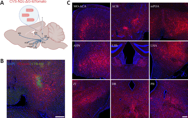
The authors first examined long-range retrograde labeling by N2cG-coated CVS-N2c-ΔG in adult C57BL/6 mice. After co-injecting tdTomato-expressing virus with green CTB-488 dye to mark the ventral tegmental area (VTA) injection site, clear local transduction was accompanied by dense retrograde labeling across multiple upstream regions known to project to the VTA.
Histological sections showed retrogradely labeled neurons distributed across somatomotor and anterior cingulate cortices, nucleus accumbens, medial preoptic area, anterior hypothalamic nucleus, lateral habenula, lateral hypothalamic area, zona incerta, dorsal raphe nucleus and parabrachial nucleus, confirming efficient axon-terminal uptake across diverse afferent populations.
Source: Lin KZ et al. · Neural regeneration research · CC BY-NC-SA
Original figure and English caption ↗
Co-injection in dorsolateral striatum yields sixfold higher VTA/SNc neuron counts than rAAV9-Retro
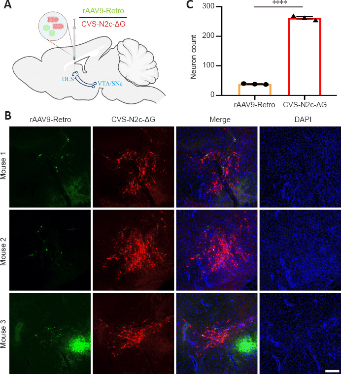
To test performance directly in a circuit where retrograde AAV vectors show limited efficiency, the team targeted the projection from the VTA and substantia nigra pars compacta (VTA/SNc) to the dorsolateral striatum (DLS). N2cG-coated CVS-N2c-ΔG-tdTomato and rAAV9-Retro-CAG-EGFP were mixed and co-injected into the DLS of adult mice.
Within the VTA/SNc, rAAV9-Retro produced only sparse EGFP-positive cells (38.33 ± 0.88 neurons), whereas N2cG-coated CVS-N2c-ΔG labeled 262.30 ± 4.06 tdTomato-positive projection neurons—roughly six times as many cells in the same pathway (P < 0.0001). This within-animal comparison demonstrates robust retrograde access to nigrostriatal and mesostriatal projection neurons that remain under-sampled by rAAV9-Retro.
Source: Lin KZ et al. · Neural regeneration research · CC BY-NC-SA
Original figure and English caption ↗
With oG complementation, contralateral hippocampal convergence triples that of SAD-B19-ΔG
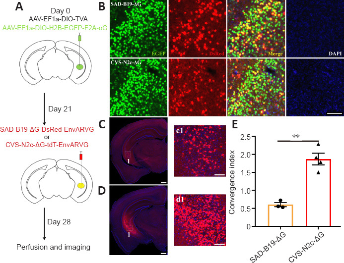
In cell-type-specific retrograde monosynaptic tracing, the pairing of viral strain and helper glycoprotein shapes how thoroughly upstream inputs are captured. After verifying in wild-type mice that EnvARVG-pseudotyped rabies vectors produced no local leak fluorescence without TVA, the authors delivered Cre-dependent AAV helpers expressing TVA and codon-optimized Pasteur-strain glycoprotein (oG) into the ventral hippocampus (vHPC) of Thy1-Cre mice, followed 3 weeks later by EnvARVG-pseudotyped CVS-N2c-ΔG or SAD-B19-ΔG.
Quantified by the convergence index—the ratio of contralateral vHPC input neurons to local GFP+dsRed+ starter cells—oG-mediated CVS-N2c-ΔG reached 1.87 ± 0.16, roughly three times the efficiency of oG-mediated SAD-B19-ΔG (0.60 ± 0.06; P = 0.0014). Additional helper comparisons showed equivalent contralateral vHPC convergence between oG and native N2cG complementation (1.87 ± 0.16 vs. 1.95 ± 0.15; P = 0.7337), and no significant difference between N2cG and codon-modified optiG across cortical, basolateral amygdalar and thalamic inputs to the striatum in D2R-Cre mice.
Source: Lin KZ et al. · Neural regeneration research · CC BY-NC-SA
Original figure and English caption ↗
Retrograde GCaMP6s delivery tracks sucrose-reward calcium transients up to 21 days
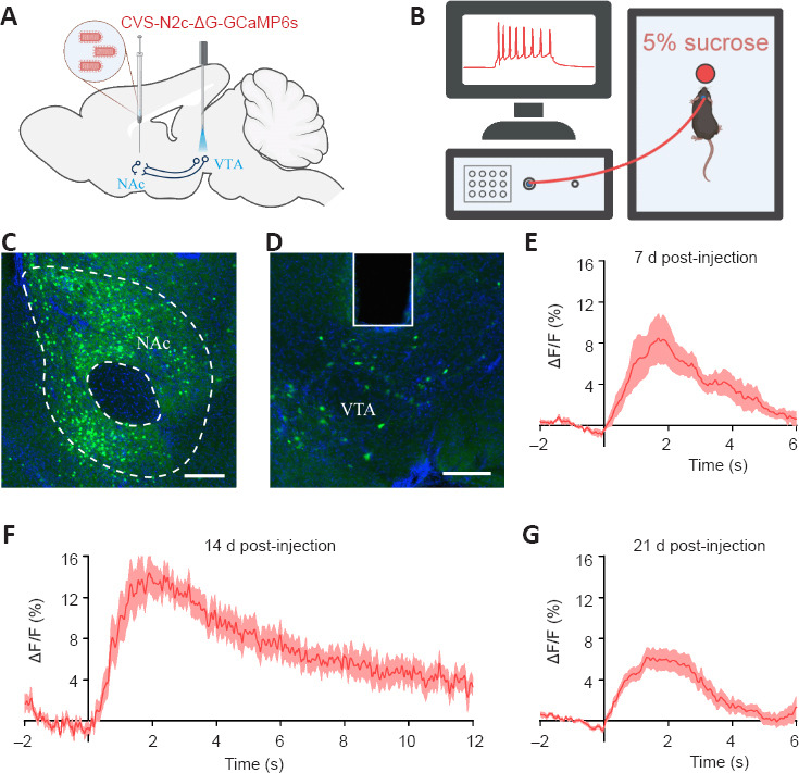
Beyond anatomical mapping, the study tested whether N2cG-coated CVS-N2c-ΔG preserves neuronal physiology long enough for longitudinal in vivo activity recording. The authors injected N2cG-coated CVS-N2c-ΔG-GCaMP6s into the nucleus accumbens (NAc) and implanted an optical fiber in the upstream VTA to monitor projection-specific calcium transients while mice consumed a 5% sucrose reward.
At 7, 14 and 21 days post-injection, sucrose consumption consistently evoked clear ΔF/F calcium elevations in retrogradely labeled VTA projection neurons. Thus, the vector supports both retrograde pathway targeting and functional monitoring of reward-evoked neural activity across at least a 21-day window.
Source: Lin KZ et al. · Neural regeneration research · CC BY-NC-SA
Original figure and English caption ↗
Efficient Cre and Flpo recombination in callosal projections alongside remaining toxicity limits
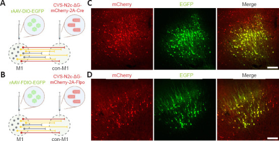
To test recombinase-mediated circuit manipulation, the authors injected Cre-dependent (rAAV-DIO-EGFP) or Flpo-dependent (rAAV-FDIO-EGFP) reporter AAVs into primary motor cortex (M1) and delivered N2cG-coated CVS-N2c-ΔG carrying mCherry-2A-Cre or mCherry-2A-Flpo into contralateral M1. Both vectors retrogradely transduced callosal projection neurons and drove robust EGFP co-labeling with mCherry in M1.
Together, the CVS-N2c-ΔG toolkit combines strong retrograde access in pathways under-labeled by rAAV9-Retro, high-convergence monosynaptic tracing with oG or N2cG, a 21-day window for in vivo calcium photometry and reliable Cre/Flpo recombination. As the authors note, although neuronal toxicity is lower than with SAD-B19-ΔG and permits three-week functional monitoring, residual toxicity is not completely eliminated and remains a target for further attenuation in longer-term studies.
Source: Lin KZ et al. · Neural regeneration research · CC BY-NC-SA
Original figure and English caption ↗